Optische Kohärenztomographie (OCT)
Anwendung und Entwicklung von ultraschnellen MHz-OCT-Systemen
Die OCT ist ein nichtinvasives Bildgebungsverfahren, welches man typischerweise nutzt um dreidimensionale Tomogramme mit hoher Auflösung (~10µm) von stark streuendem Gewebe zu erstellen. Durch die Verwendung von eigens entwickelten FDML-Lasern erreichen wir Aufnahmegeschwindigkeiten von mehreren Millionen Tiefenscans pro Sekunde (MHz-OCT). Dies ist um ein bis zwei Größenordnungen schneller als derzeitige kommerzielle Systeme.
Diese hohen Geschwindigkeiten sind in vielen klinischen Bereichen (z.B. ophthalmisches und intravaskuläres OCT) nützlich, da sie die Aufnahmedauer verringern und helfen Bewegungsartefakte zu vermeiden. Die hohe Geschwindigkeit ermöglicht aber auch einen Zugang zur Phase des detektierten Lichts und damit neue numerische Methoden zur Bildverbesserung und Kontrastgebung in der Swept-Source-OCT.
Unsere Arbeitsgruppe forscht im Bereich der OCT an neuen Technologien und zeigt mögliche Anwendungsgebiete auf.
Forschungsschwerpunkte:
- MHz-OCT - Ultraschnelle OCT-Bildgebung mit mehreren millionen Tiefenschnitten pro Sekunde
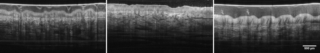
- LARA-OCT - Großflächige OCT-Bildgebung von Haut mittel Roboter unterstützer MHz-OCT
- VR-OCT - Echtzeit Berechnung und Visualisierung ganzer OCT-Volumen in einer virtuellen Umgebung
- Augen OCT - Anwendung der MHz-OCT am Auge zur Darstellung der Netzhaut oder des Augenvordergrunds
- Phasensensitive OCT - Erweiterung des Informationsgehalts einer OCT-Aufnahme durch hinzufügen eines Phasenkontrastes
- Multispektrale OCT - Kombination aus RGB- und OCT-Aufnahmen zur verbesserten Darstellung morphologischer Strukturen
zugehörige Publikationen
2021
Creating a depth-resolved OCT-dataset for supervised classification based on ex vivo human brain samples, in Optical Coherence Tomography and Coherence Domain Optical Methods in Biomedicine XXV , SPIE, Mä.2021. pp. 66 -- 73.
| DOI: | 10.1117/12.2578391 |
| Bibtex: | @inproceedings{Strenge2021,
author = {P. Strenge, B. Lange, C. Grill, W. Draxinger, V. Danicke, D. Theisen-Kunde, H. Handels, C. Hagel, M. Bonsanto, R. Huber and R. Brinkmann},
title = {{Creating a depth-resolved OCT-dataset for supervised classification based on ex vivo human brain samples}},
volume = {11630},
booktitle = {Optical Coherence Tomography and Coherence Domain Optical Methods in Biomedicine XXV},
editor = {Joseph A. Izatt and James G. Fujimoto},
organization = {International Society for Optics and Photonics},
publisher = {SPIE},
pages = {66 -- 73},
abstract = {Optical coherence tomography (OCT) has the potential to become an additional imaging modality for surgical guidance in the field of neurosurgery, especially when it comes to the detection of different infiltration grades of glioblastoma multiforme at the tumor border. Interpretation of the images, however, is still a big challenge. A method to create a labeled OCT dataset based on ex vivo brain samples is introduced. The tissue samples were embedded in an agarose mold giving them a distinctive shape before images were acquired with two OCT systems (spectral domain (SD) and swept source (SS) OCT) and histological sections were created and segmented by a neuropathologist. Based on the given shape, the corresponding OCT images for each histological image can be determined. The transfer of the labels from the histological images onto the OCT images was done with a non-affine image registration approach based on the tissue shape. It was demonstrated that finding OCT images of a tissue sample corresponding to segmented histological images without any color or laser marking is possible. It was also shown that the set labels can be transferred onto OCT images. The accuracy of method is 26 ± 11 pixel, which translates to 192 ± 75 μm for the SS-OCT and 94 ± 43 μm for the SD-OCT. The dataset consists of several hundred labeled OCT images, which can be used to train a classification algorithm.},
keywords = {AG-Huber_OCT, optical coherence tomography, OCT, image registration, glioblastoma multiforme, MHz-OCT, brain imaging, tumor, neurosurgery},
year = {2021},
URL = {https://doi.org/10.1117/12.2578391}
} |
1.6 MHz FDML OCT for Intraoperative Imaging in Neurosurgery, in European Conferences on Biomedical Optics 2021 (ECBO) , Optica Publishing Group, 2021. pp. ETu4A.2.
| Weblink: | https://opg.optica.org/abstract.cfm?URI=ECBO-2021-ETu4A.2 |
| Datei: | abstract.cfm |
| Bibtex: | @inproceedings{Theisen-Kunde:21,
author = {D. Theisen-Kunde and W. Draxinger and M. M. Bonsanto and Paul Strenge and Nicolas Detrez and R. Huber and R. Brinkmann},
booktitle = {European Conferences on Biomedical Optics 2021 (ECBO)},
journal = {European Conferences on Biomedical Optics 2021 (ECBO)},
keywords = {Clinical applications; Fourier domain mode locking; Optical coherence tomography; Optical fibers; Three dimensional reconstruction; White light},
pages = {ETu4A.2},
publisher = {Optica Publishing Group},
title = {1.6 MHz FDML OCT for Intraoperative Imaging in Neurosurgery},
year = {2021},
url = {https://opg.optica.org/abstract.cfm?URI=ECBO-2021-ETu4A.2},
doi = {10.1364/ECBO.2021.ETu4A.2},
abstract = {A 1.6 MHz Fourier-domain mode-locked (FDML) optical coherence tomography (OCT) was adapted to an OR-Microscope for clinical application in neurosurgery. 3D-volume scans at video rate are envisaged with approximately 50{\textmu}m lateral and 20{\textmu}m axial resolution.},
} |
Characterization of the dynamics of an FDML laser during closed-loop cavity length control, in Fiber Lasers XVIII: Technology and Systems , Michalis N. Zervas, Eds. SPIE, 2021. pp. 236 -- 241.
| DOI: | 10.1117/12.2578514 |
| Bibtex: | @inproceedings{LotzLASE2021,
author = {S. Lotz, C. Grill, M. Göb, W. Draxinger, J. P. Kolb and R. Huber},
title = {{Characterization of the dynamics of an FDML laser during closed-loop cavity length control}},
volume = {11665},
booktitle = {Fiber Lasers XVIII: Technology and Systems},
editor = {Michalis N. Zervas},
organization = {International Society for Optics and Photonics},
publisher = {SPIE},
pages = {236 -- 241},
abstract = {In Fourier domain mode locked (FDML) lasers, extremely precise and stable matching of the filter tuning period and light circulation time in the cavity is essential for ultra-low noise operation. During the operation of FDML lasers, the ultra-low noise mode can be lost due to temperature drifts of the already temperature stabilized cavity resulting in increased intensity noise. Until now, the filter frequency is continuously regulated to match the changing light circulation time. However, this causes the filter frequency to constantly change by a few mHz and leads to synchronization issues in cases where a fixed filter frequency is desired. We present an actively cavity length controlled FDML laser and a robust and high precision feedback loop algorithm for maintaining ultra-low noise operation. Instead of adapting the filter frequency, the cavity length is adjusted by a motorized free space beam path to match the fixed filter frequency. The closed-loop system achieves a stability of ~0.18 mHz at a sweep repetition rate of ~418 kHz which corresponds to a ratio of 4×10<sup>-10</sup>. We investigate the coherence properties during the active cavity length adjustments and observe no noise increase compared to fixed cavity length. The cavity length control is fully functional and for the first time, offers the possibility to operate an FDML laser in sweet spot mode at a fixed frequency or phase locked to an external clock. This opens new possibilities for system integration of FDML lasers.},
keywords = {AG-Huber_FDML, FDML, Fourier domain mode locking, laser beating, tunable laser, optical coherence tomography, OCT},
year = {2021},
URL = {hhttps://doi.org/10.1117/12.2578514}
} |
2020
Flexible A-scan rate MHz-OCT: efficient computational downscaling by coherent averaging, Biomed. Opt. Express , vol. 11, no. 11, pp. 6799--6811, Nov. 2020. OSA.
| DOI: | 10.1364/BOE.402477 |
| Bibtex: | @article{Pfeiffer:20,
author = {T. Pfeiffer, M. G\"{o}b, W. Draxinger, S. Karpf, J.P. Kolb and R. Huber},
journal = {Biomed. Opt. Express},
keywords = {AG-Huber_OCT; High speed imaging; Image quality; Optical coherence tomography; Swept lasers; Swept sources; Systems design},
number = {11},
pages = {6799--6811},
publisher = {OSA},
title = {Flexible A-scan rate MHz-OCT: efficient computational downscaling by coherent averaging},
volume = {11},
month = {Nov},
year = {2020},
doi = {10.1364/BOE.402477},
abstract = {In order to realize adjustable A-scan rates of fast optical coherence tomography (OCT) systems, we investigate averaging of OCT image data acquired with a MHz-OCT system based on a Fourier Domain Mode Locked (FDML) laser. Increased system sensitivity and image quality can be achieved with the same system at the cost of lower imaging speed. Effectively, the A-scan rate can be reduced in software by a freely selectable factor. We demonstrate a detailed technical layout of the strategies necessary to achieve efficient coherent averaging. Since there are many new challenges specific to coherent averaging in swept source MHz-OCT, we analyze them point by point and describe the appropriate solutions. We prove that coherent averaging is possible at MHz OCT-speed without special interferometer designs or digital phase stabilization. We find, that in our system up to \&\#x223C;100x coherent averaging is possible while achieving a sensitivity increase close to the ideal values. This corresponds to a speed reduction from 3.3 MHz to 33 kHz and a sensitivity gain of 20 dB. We show an imaging comparison between coherent and magnitude averaging of a human finger knuckle joint in vivo with 121\&\#x00A0;dB sensitivity for the coherent case. Further, the benefits of computational downscaling in low sensitivity MHz-OCT systems are analyzed.},
}
|
Beating of two FDML lasers in real time, in Fiber Lasers XVII: Technology and Systems , Liang Dong, Eds. SPIE, Feb.2020. pp. 132 -- 138.
| DOI: | 10.1117/12.2545794 |
| Bibtex: | @inproceedings{Grill2020,
author = {C. {Grill}, S. {Lotz}, T. {Blömker}, D. {Kastner}, T. {Pfeiffer}, S. {Karpf}, M. {Schmidt}, W. {Draxinger}, C.
{Jirauschek} and R. {Huber}},
title = {{Beating of two FDML lasers in real time}},
volume = {11260},
booktitle = {Fiber Lasers XVII: Technology and Systems},
editor = {Liang Dong},
organization = {International Society for Optics and Photonics},
publisher = {SPIE},
pages = {132 -- 138},
keywords = {AG-Huber_FDML, FDML laser, fiber lasers, beat signal, OCT, Optical Coherence Tomography, Fourier domain mode locking},
year = {2020},
doi = {10.1117/12.2545794},
}
|
Segmented OCT data set for depth resolved brain tumor detection validated by histological analysis, in Optical Coherence Tomography and Coherence Domain Optical Methods in Biomedicine XXIV , SPIE, Feb.2020. pp. 82 -- 89.
| DOI: | 10.1117/12.2545659 |
| Bibtex: | @inproceedings{Strenge2020,
author = {P. Strenge and B. Lange and C. Grill and W. Draxinger and M. M. Bonsanto and C. Hagel and R. Huber and R. Brinkmann},
title = {{Segmented OCT data set for depth resolved brain tumor detection validated by histological analysis}},
volume = {11228},
booktitle = {Optical Coherence Tomography and Coherence Domain Optical Methods in Biomedicine XXIV},
editor = {Joseph A. Izatt and James G. Fujimoto},
organization = {International Society for Optics and Photonics},
publisher = {SPIE},
pages = {82 -- 89},
keywords = {AG-Huber_OCT, Optical coherence tomography, OCT, FDML Laser, MHz-OCT, brain tumor, brain imaging, neurosurgery},
year = {2020},
URL = { https://www.spiedigitallibrary.org/conference-proceedings-of-spie/11228/112282O/Segmented-OCT-data-set-for-depth-resolved-brain-tumor-detection/10.1117/12.2545659.short}
}
|
In-vitro and in-vivo imaging of coronary artery stents with Heartbeat OCT, The International Journal of Cardiovascular Imaging , vol. 36, no. 6, pp. 1021-1029, Feb. 2020. Springer Science and Business Media LLC.
| DOI: | 10.1007/s10554-020-01796-7 |
| Bibtex: | @article{Cecchetti2020,
doi = {10.1007/s10554-020-01796-7},
url = {https://doi.org/10.1007/s10554-020-01796-7},
year = {2020},
month = feb,
publisher = {Springer Science and Business Media {LLC}},
volume = {36},
number = {6},
pages = {1021--1029},
author = {Leonardo Cecchetti and Tianshi Wang and Ayla Hoogendoorn and Karen T. Witberg and Jurgen M. R. Ligthart and Joost Daemen and Heleen M. M. van Beusekom and Tom Pfeiffer and Robert A. Huber and Jolanda J. Wentzel and Antonius F. W. van der Steen and Gijs van Soest},
title = {In-vitro and in-vivo imaging of coronary artery stents with Heartbeat {OCT}},
journal = {The International Journal of Cardiovascular Imaging}
} |
2019
Motorized capsule for shadow-free OCT imaging and synchronous beam control, Opt Lett , vol. 44, no. 15, pp. 3641-3644, Aug. 2019. Optica Publishing Group.
| DOI: | 10.1364/OL.44.003641 |
| Bibtex: | @article{Lopez-Marin:19,
author = {Antonio L\'{o}pez-Mar\'{i}n and Geert Springeling and Robert Beurskens and Heleen van Beusekom and Antonius F. W. van der Steen and Arjun D. Koch and Brett E. Bouma and Robert Huber and Gijs van Soest and Tianshi Wang},
journal = {Opt. Lett.},
keywords = {Image reconstruction; Light beams; Magnetic fields; Optical coherence tomography; Optical imaging; Reflector design},
number = {15},
pages = {3641--3644},
publisher = {Optica Publishing Group},
title = {Motorized capsule for shadow-free OCT imaging and synchronous beam control},
volume = {44},
month = {Aug},
year = {2019},
url = {https://opg.optica.org/ol/abstract.cfm?URI=ol-44-15-3641},
doi = {10.1364/OL.44.003641},
abstract = {We demonstrate a tethered motorized capsule for unobstructed optical coherence tomography (OCT) imaging of the esophagus. By using a distal reflector design, we avoided the common shadow artifact induced by the motor wires. A synchronous driving technique features three types of beam-scanning modes of the capsule, i.e., circumferential beam scanning, localized beam scanning, and accurate beam positioning. We characterized these three modes and carried out ex vivo imaging experiments using the capsule. The results show that the capsule can potentially be a useful tool for diagnostic OCT imaging and OCT-guided biopsy and therapy of the esophagus.},
} |
MHz-OCT for low latency virtual reality guided surgery: first wet lab experiments on ex-vivo porcine eye, in Optical Coherence Imaging Techniques and Imaging in Scattering Media III , Maciej Wojtkowski and Stephen A. Boppart and Wang-Yuhl Oh, Eds. SPIE, Jul.2019. pp. 110780E.
| DOI: | 10.1117/12.2527123 |
| Bibtex: | @inproceedings{10.1117/12.2527123,
author = {Yoko Miura and Wolfgang Draxinger and Christin Grill and Tom Pfeiffer and Salvatore Grisanti and Robert Huber},
title = {{MHz-OCT for low latency virtual reality guided surgery: first wet lab experiments on ex-vivo porcine eye
}},
volume = {11078},
booktitle = {Optical Coherence Imaging Techniques and Imaging in Scattering Media III},
editor = {Maciej Wojtkowski and Stephen A. Boppart and Wang-Yuhl Oh},
organization = {International Society for Optics and Photonics},
publisher = {SPIE},
pages = {110780E},
abstract = {MHz-OCT systems based on FDML swept laser sources combined with the massive parallel processing capabilities of modern computer hardware enable volumetric imaging, processing and stereoscopic display at video rates. The increasing image quality and speed might enable new fields of application where the volumetric OCT completely replaces stereoscopic microscopes instead of being a mere supplement. Aside from the depth resolving capability, a particular advantage is the ability to display a whole image volume from arbitrary points of view without the need to move the actual microscope or to rotate the patient’s eye. Purely digital microscopy is already offered as alternative to traditional through-an-eyepiece surgical microscope. We explore the use of virtual reality to present digital OCT microscopy images to a trained surgeon, carrying out a series of surgical procedures ex-vivo on a porcine eye model.},
keywords = {virtual reality, surgery guidance , real-time OCT, user experience},
year = {2019},
doi = {10.1117/12.2527123},
URL = {https://doi.org/10.1117/12.2527123}
} |
Towards combined optical coherence tomography and multi-spectral imaging with MHz a-scan rates for endoscopy, in Optical Coherence Imaging Techniques and Imaging in Scattering Media III , aciej Wojtkowski and Stephen A. Boppart and Wang-Yuhl Oh, Eds. Jul.2019. pp. 110780Y.
| DOI: | 10.1117/12.2526796 |
| Bibtex: | @inproceedings{10.1117/12.2526796,
author = {Madita G{\"o}b and Tom Pfeiffer and Robert Huber},
title = {{Towards combined optical coherence tomography and multi-spectral imaging with MHz a-scan rates for endoscopy}},
volume = {11078},
booktitle = {Optical Coherence Imaging Techniques and Imaging in Scattering Media III},
editor = {Maciej Wojtkowski and Stephen A. Boppart and Wang-Yuhl Oh},
organization = {International Society for Optics and Photonics},
publisher = {SPIE},
pages = {110780Y},
abstract = {We demonstrate a preliminary setup of a combined MHz-OCT and RGB narrowband reflection microscope and investigate the performance of the new RGB branch and different display modes of colored OCT data sets.},
keywords = {MHz OCT, multi-spectral imaging, Optical Coherence Tomography, Fourier Domain Mode Locked , FDML, RGB, Color },
year = {2019},
doi = {10.1117/12.2526796},
URL = {https://doi.org/10.1117/12.2526796}
}
|
Zero roll-off retinal MHz-OCT using an FDML-laser, in Optical Coherence Imaging Techniques and Imaging in Scattering Media III , SPIE, Jul.2019. pp. 110780S.
| DOI: | 10.1117/12.2527034 |
| Datei: | 12.2527034.short |
| Bibtex: | @inproceedings{10.1117/12.2527034,
author = {Julian Klee and Jan Philip Kolb and Christin Grill and Wolfgang Draxinger and Tom Pfeiffer and Robert Huber},
title = {{Zero roll-off retinal MHz-OCT using an FDML-laser}},
volume = {11078},
booktitle = {Optical Coherence Imaging Techniques and Imaging in Scattering Media III},
editor = {Maciej Wojtkowski and Stephen A. Boppart and Wang-Yuhl Oh},
organization = {International Society for Optics and Photonics},
publisher = {SPIE},
pages = {110780S},
abstract = {Optical coherence tomography (OCT) applications like ultra-widefield and full eye-length imaging are of high interest for various diagnostic purposes. In swept-source OCT these techniques require a swept light source, which is coherent over the whole imaging depth. We present a zero roll-off 1060 nm Fourier Domain Mode Locked-Laser (FDML-Laser) for retinal OCT imaging at 1.7 MHz A-scan rate and first long-range imaging results with it. Several steps such as improved dispersion compensation and frequency regulation were performed and will be discussed. Besides virtually no loss in OCT signal over the maximum depth range of 4.6 mm and very good dynamic range was observed. Roll-off measurements show no decrease of the point-spread function (PSF), while maintaining a high dynamic range.},
keywords = {optical coherence tomography, OCT, tunable laser, Fourier Domain Mode Locking, FDML, MHz OCT},
year = {2019},
doi = {10.1117/12.2527034},
URL = {https://doi.org/10.1117/12.2527034}
} |
Live video rate volumetric OCT imaging of the retina with multi-MHz A-scan rates, PLOS ONE , vol. 14, no. 7, pp. e0213144, Mä. 2019.
| DOI: | 10.1371/journal.pone.0213144 |
| Bibtex: | @article{Kolb2019,
author = {Kolb, J P;Draxinger, W;Klee, J;Pfeiffer, T;Eibl, M;Klein, T;Wieser, W and Huber, R},
title = {Live video rate volumetric OCT imaging of the retina with multi-MHz A-scan rates},
journal = {J pone},
keywords = {AG-Huber_OCT},
url = {https://doi.org/10.1371/journal.pone.0213144},
pages = {e0213144},
ISSN = {1932-6203},
year = {2019},
type = {Journal Article}
}
|
Measurement of Inter-Sweep Phase Stability of an FDML Laser with a 10 kHz Tunable Ring Laser, in 2019 Conference on Lasers and Electro-Optics Europe and European Quantum Electronics Conference , Optical Society of America, 2019. pp. 1-1.
| DOI: | 10.1109/CLEOE-EQEC.2019.8872860 |
| Bibtex: | @inproceedings{Kastner:19,
author = {Kastner, D; Bl\"{o}mker, T; Pfeiffer, T; Grill, C; Schmidt, M; Jirauschek, C and Huber, R},
booktitle = {2019 Conference on Lasers and Electro-Optics Europe and European Quantum Electronics Conference},
journal = {2019 Conference on Lasers and Electro-Optics Europe and European Quantum Electronics Conference},
keywords = {Fourier domain mode locking; Image quality; Optical coherence tomography; Phase noise; Ring lasers; Tunable lasers},
pages = {cj_7_5},
publisher = {Optical Society of America},
title = {Measurement of Inter-Sweep Phase Stability of an FDML Laser with a 10 kHz Tunable Ring Laser},
year = {2019},
keywords = {AG-Huber_FDML, AG-Huber_OCT},
doi = { 10.1109/CLEOE-EQEC.2019.8872860},
abstract = {Fourier Domain Mode Locking (FDML) lasers are light sources that generate a sequence of narrowband optical frequency sweeps at the fundamental or harmonic of the cavity repetition rate \[1\]. This frequency swept output can also be considered as a sequence of strongly chirped, long pulses. FDML lasers are mainly used in swept source optical coherence tomography (SS-OCT), a medical imaging technique. The coherence length of the source, i.e. the intra-sweep phase stability of an FDML sweep, is decisive for the image quality and performance of OCT imaging \[2\].},
} |
2018
Combined in-depth, 3D, en face imaging of the optic disc, optic disc pits and optic disc pit maculopathy using swept-source megahertz OCT at 1050 nm, Graefes Arch Clin Exp Ophthalmol , vol. 256, no. 2, pp. 289-298, Dez. 2018.
| DOI: | 10.1007/s00417-017-3857-9 |
| Bibtex: | @article{Maertz2018,
author = {Maertz, J; Kolb, J P; Klein, T; Mohler, K J; Eibl, M; Wieser, W; Huber, R; Priglinger, S and Wolf, A},
title = {Combined in-depth, 3D, en face imaging of the optic disc, optic disc pits and optic disc pit maculopathy using swept-source megahertz OCT at 1050 nm},
journal = {Graefe's Archive for Clinical and Experimental Ophthalmology},
number = {2},
pages = {289-298},
DOI = {10.1007/s00417-017-3857-9},
url = {https://www.scopus.com/inward/record.uri?eid=2-s2.0-85032262413&doi=10.1007%2fs00417-017-3857-9&partnerID=40&md5=a46c315f12cf5e633ea0f7e644116eb3},
year = {2018},
Keywords= {En face imaging, Optical coherence tomography, Swept-source OCT, Megahertz OCT, 3D rendering, Optic disc, Optic disc pit, Optic disc pit maculopathy, AG-Huber_OCT},
type = {Journal Article}
} |
High-speed fiber scanning endoscope for volumetric multi-megahertz optical coherence tomography, Opt. Lett. , vol. 43, no. 18, pp. 4386-4389, Sep. 2018. Optica Publishing Group.
| DOI: | 10.1364/OL.43.004386 |
| Bibtex: | @article{Schulz-Hildebrandt:18,
author = {Hinnerk Schulz-Hildebrandt and Tom Pfeiffer and Tim Eixmann and Sabrina Lohmann and Martin Ahrens and Joshua Rehra and Wolfgang Draxinger and Peter K\"{o}nig and Robert Huber and Gereon H\"{u}ttmann},
journal = {Opt. Lett.},
keywords = {Fiber optics imaging; Endoscopic imaging; Medical and biological imaging; Optical coherence tomography; Fourier domain mode locking; Image quality; Optical coherence tomography; Single mode fibers; Step index fibers; Three dimensional imaging},
number = {18},
pages = {4386--4389},
publisher = {Optica Publishing Group},
title = {High-speed fiber scanning endoscope for volumetric multi-megahertz optical coherence tomography},
volume = {43},
month = {Sep},
year = {2018},
url = {https://opg.optica.org/ol/abstract.cfm?URI=ol-43-18-4386},
doi = {10.1364/OL.43.004386},
abstract = {We present a forward-viewing fiber scanning endoscope (FSE) for high-speed volumetric optical coherence tomography (OCT). The reduction in size of the probe was achieved by substituting the focusing optics by an all-fiber-based imaging system which consists of a combination of scanning single-mode fibers, a glass spacer, made from a step-index multi-mode fiber, and a gradient-index fiber. A lateral resolution of 11 $\mu$m was achieved at a working distance of 1.2 mm. The newly designed piezo-based FSE has an outer diameter of 1.6 mm and a rigid length of 13.5 mm. By moving the whole imaging optic in spirals for scanning the sample, the beam quality remains constant over the entire field of view with a diameter of 0.8 mm. The scanning frequency was adjusted to 1.22 kHz for use with a 3.28 MHz Fourier domain mode locked OCT system. Densely sampled volumes have been imaged at a rate of 6 volumes per second.},
} |
Ultra low noise Fourier domain mode locked laser for high quality magahertz optical coherence tomography, Biomed. Opt. Express , vol. 9, no. 9, pp. 4130-4148, Sep. 2018. Optica Publishing Group.
| DOI: | 10.1364/BOE.9.004130 |
| Bibtex: | @article{Pfeiffer:18,
author = {Tom Pfeiffer and Markus Petermann and Wolfgang Draxinger and Christian Jirauschek and Robert Huber},
journal = {Biomed. Opt. Express},
keywords = {Fiber optics imaging; Lasers, fiber; Optical coherence tomography; Laser stabilization ; Lasers, frequency modulated ; Analog to digital converters; Dark solitons; Image quality; Laser modes; Mode locking; Optical coherence tomography},
number = {9},
pages = {4130--4148},
publisher = {Optica Publishing Group},
title = {Ultra low noise Fourier domain mode locked laser for high quality megahertz optical coherence tomography},
volume = {9},
month = {Sep},
year = {2018},
url = {https://opg.optica.org/boe/abstract.cfm?URI=boe-9-9-4130},
doi = {10.1364/BOE.9.004130},
abstract = {We investigate the origin of high frequency noise in Fourier domain mode locked (FDML) lasers and present an extremely well dispersion compensated setup which virtually eliminates intensity noise and dramatically improves coherence properties. We show optical coherence tomography (OCT) imaging at 3.2 MHz A-scan rate and demonstrate the positive impact of the described improvements on the image quality. Especially in highly scattering samples, at specular reflections and for strong signals at large depth, the noise in optical coherence tomography images is significantly reduced. We also describe a simple model that suggests a passive physical stabilizing mechanism that leads to an automatic compensation of remaining cavity dispersion in FDML lasers.},
} |
High-resolution retinal swept source optical coherence tomography with an ultra-wideband Fourier-domain mode-locked laser at MHz A-scan rates, Biomed. Opt. Express , vol. 9, no. 1, pp. 120-130, Jan. 2018. Optica Publishing Group.
| DOI: | 10.1364/BOE.9.000120 |
| Bibtex: | @article{Kolb:18,
author = {Jan Philip Kolb and Tom Pfeiffer and Matthias Eibl and Hubertus Hakert and Robert Huber},
journal = {Biomed. Opt. Express},
keywords = {Medical optics instrumentation; Lasers, fiber; Medical and biological imaging; Ophthalmic optics and devices ; Optical coherence tomography; Adaptive optics; Image quality; In vivo imaging; Mode locking; Ophthalmic imaging; Three dimensional imaging},
number = {1},
pages = {120--130},
publisher = {Optica Publishing Group},
title = {High-resolution retinal swept source optical coherence tomography with an ultra-wideband Fourier-domain mode-locked laser at MHz A-scan rates},
volume = {9},
month = {Jan},
year = {2018},
url = {https://opg.optica.org/boe/abstract.cfm?URI=boe-9-1-120},
doi = {10.1364/BOE.9.000120},
abstract = {We present a new 1060 nm Fourier domain mode locked laser (FDML laser) with a record 143 nm sweep bandwidth at 2\&\#x2219;\&\#x202F;417 kHz\&\#x202F; $=$ \&\#x202F;834 kHz and 120 nm at 1.67 MHz, respectively. We show that not only the bandwidth alone, but also the shape of the spectrum is critical for the resulting axial resolution, because of the specific wavelength-dependent absorption of the vitreous. The theoretical limit of our setup lies at 5.9 \&\#x00B5;m axial resolution. In vivo MHz-OCT imaging of human retina is performed and the image quality is compared to the previous results acquired with 70 nm sweep range, as well as to existing spectral domain OCT data with 2.1 \&\#x00B5;m axial resolution from literature. We identify benefits of the higher resolution, for example the improved visualization of small blood vessels in the retina besides several others.},
} |
2017
Thermo-elastic optical coherence tomography, Optica Publishing Group, Sep.2017. pp. 3466-3469.
| DOI: | 10.1364/OL.42.003466 |
| Bibtex: | @article{Wang:17,
author = {Tianshi Wang and Tom Pfeiffer and Min Wu and Wolfgang Wieser and Gaetano Amenta and Wolfgang Draxinger and Antonius F. W. van der Steen and Robert Huber and Gijs van Soest},
journal = {Opt. Lett.},
keywords = {Imaging systems; Medical and biological imaging; Optical coherence tomography; Lasers, pulsed ; Fourier domain mode locking; Functional imaging; Laser beams; Nanosecond pulses; Optical coherence tomography; Phantom studies},
number = {17},
pages = {3466--3469},
publisher = {Optica Publishing Group},
title = {Thermo-elastic optical coherence tomography},
volume = {42},
month = {Sep},
year = {2017},
url = {https://opg.optica.org/ol/abstract.cfm?URI=ol-42-17-3466},
doi = {10.1364/OL.42.003466},
abstract = {The absorption of nanosecond laser pulses induces rapid thermo-elastic deformation in tissue. A sub-micrometer scale displacement occurs within a few microseconds after the pulse arrival. In this Letter, we investigate the laser-induced thermo-elastic deformation using a 1.5 MHz phase-sensitive optical coherence tomography (OCT) system. A displacement image can be reconstructed, which enables a new modality of phase-sensitive OCT, called thermo-elastic OCT. An analysis of the results shows that the optical absorption is a dominating factor for the displacement. Thermo-elastic OCT is capable of visualizing inclusions that do not appear on the structural OCT image, providing additional tissue type information.},
} |
1060nm FDML laser with centimeter coherence length and 1.67 MHz sweep rate for full eye length and retinal ultra-widefield OCT, in Optical Coherence Imaging Techniques and Imaging in Scattering Media II , Maciej Wojtkowski and Stephen A. Boppart and Wang-Yuhl Oh, Eds. SPIE, Aug.2017. pp. 104160J.
| DOI: | 10.1117/12.2286854 |
| Bibtex: | @inproceedings{10.1117/12.2286854,
author = {Jan Philip Kolb and Julian Klee and Tom Pfeiffer and Robert Huber},
title = {{1060nm FDML laser with centimeter coherence length and 1.67 MHz sweep rate for full eye length and retinal ultra-widefield OCT}},
volume = {10416},
booktitle = {Optical Coherence Imaging Techniques and Imaging in Scattering Media II},
editor = {Maciej Wojtkowski and Stephen A. Boppart and Wang-Yuhl Oh},
organization = {International Society for Optics and Photonics},
publisher = {SPIE},
pages = {104160J},
abstract = {We present a new design of a 1060nm Fourier Domain Mode Locked-Laser (FDML-Laser) that combines 1.67 MHz A-scan rate with a centimeter scale coherence length. The extended coherence length is achieved by synchronizing the cavity roundtrip time over the 75 nm sweep with a relative accuracy of 10<sup>-7</sup>. We will show that this requires careful combination of multiple fiber types in the cavity with a gradient heated chirped Fiber Bragg grating.},
keywords = {optical coherence tomograhy, OCT, tunable laser, Fourier domain mode locking, FDML, MHz OCT},
year = {2017},
doi = {10.1117/12.2286854},
URL = {https://doi.org/10.1117/12.2286854}
}
|
Long-range live 3D-OCT at different spectral zoom levels, in Optical Coherence Imaging Techniques and Imaging in Scattering Media II , Maciej Wojtkowski and Stephen A. Boppart and Wang-Yuhl Oh, Eds. SPIE, Aug.2017. pp. 104160L.
| DOI: | 10.1117/12.2287484 |
| Bibtex: | @inproceedings{10.1117/12.2287484,
author = {Tom Pfeiffer and Wolfgang Draxinger and Christin Grill and Robert Huber},
title = {{Long-range live 3D-OCT at different spectral zoom levels}},
volume = {10416},
booktitle = {Optical Coherence Imaging Techniques and Imaging in Scattering Media II},
editor = {Maciej Wojtkowski and Stephen A. Boppart and Wang-Yuhl Oh},
organization = {International Society for Optics and Photonics},
publisher = {SPIE},
pages = {104160L},
abstract = {We demonstrate that the 3.2 MHz a-scan rate and the improved coherence of our new low noise FDML laser enables live 3D-OCT with different spectral zooms and up to 10 cm of imaging range.},
keywords = {Optical coherence tomography, Fourier Domain Mode Locking, FDML, OCT},
year = {2017},
doi = {10.1117/12.2287484},
URL = {https://doi.org/10.1117/12.2287484}
}
|
INTRAPAPILLARY PROLIFERATION IN OPTIC DISK PITS: Clinical Findings and Time-Related Changes, Retina , vol. 37, no. 5, pp. 906-914, Mai 2017.
| DOI: | 10.1097/iae.0000000000001260 |
| Bibtex: | @article{Maertz2017,
author = {Maertz, J. and Mohler, K. J. and Kolb, J. P. and Klein, T. and Neubauer, A. and Kampik, A. and Priglinger, S. and Wieser, W. and Huber, R. and Wolf, A.},
title = {INTRAPAPILLARY PROLIFERATION IN OPTIC DISK PITS: Clinical Findings and Time-Related Changes},
journal = {Retina},
volume = {37},
number = {5},
pages = {906-914},
DOI = {10.1097/iae.0000000000001260},
year = {2017},
keywords = {AG-Huber_OCT},
type = {Journal Article}
}
|
Feature tracking for automated volume of interest stabilization on 4D-OCT images, in Medical Imaging 2017: Image-Guided Procedures, Robotic Interventions, and Modeling , Robert J. Webster III and Baowei Fei, Eds. SPIE, Mä.2017. pp. 101350W.
| DOI: | 10.1117/12.2255090 |
| Bibtex: | @inproceedings{10.1117/12.2255090,
author = {Max-Heinrich Laves and Andreas Schoob and L{\"u}der A. Kahrs and Tom Pfeiffer and Robert Huber and Tobias Ortmaier},
title = {{Feature tracking for automated volume of interest stabilization on 4D-OCT images}},
volume = {10135},
booktitle = {Medical Imaging 2017: Image-Guided Procedures, Robotic Interventions, and Modeling},
editor = {Robert J. Webster III and Baowei Fei},
organization = {International Society for Optics and Photonics},
publisher = {SPIE},
pages = {101350W},
abstract = {A common representation of volumetric medical image data is the triplanar view (TV), in which the surgeon manually selects slices showing the anatomical structure of interest. In addition to common medical imaging such as MRI or computed tomography, recent advances in the field of optical coherence tomography (OCT) have enabled live processing and volumetric rendering of four-dimensional images of the human body. Due to the region of interest undergoing motion, it is challenging for the surgeon to simultaneously keep track of an object by continuously adjusting the TV to desired slices. To select these slices in subsequent frames automatically, it is necessary to track movements of the volume of interest (VOI). This has not been addressed with respect to 4DOCT images yet. Therefore, this paper evaluates motion tracking by applying state-of-the-art tracking schemes on maximum intensity projections (MIP) of 4D-OCT images. Estimated VOI location is used to conveniently show corresponding slices and to improve the MIPs by calculating thin-slab MIPs. Tracking performances are evaluated on an in-vivo sequence of human skin, captured at 26 volumes per second. Among investigated tracking schemes, our recently presented tracking scheme for soft tissue motion provides highest accuracy with an error of under 2.2 voxels for the first 80 volumes. Object tracking on 4D-OCT images enables its use for sub-epithelial tracking of microvessels for image-guidance.},
keywords = {4D imaging, maximum intensity projection, optical coherence tomography, feature tracking},
year = {2017},
doi = {10.1117/12.2255090},
URL = {https://doi.org/10.1117/12.2255090}
}
|
Short pulse laser induced thermo-elastic deformation imaging, in Optical Interactions with Tissue and Cells XXVIII , E. Duco Jansen and Hope Thomas Beier, Eds. SPIE, Feb.2017. pp. 100620C.
| DOI: | 10.1117/12.2251502 |
| Bibtex: | @inproceedings{10.1117/12.2251502,
author = {Tianshi Wang and Tom Pfeiffer and Min Wu and Wolfgang Wieser and Wolfgang Draxinger and Antonius F. W. van der Steen and Robert Huber and Gijs van Soest},
title = {{Short pulse laser induced thermo-elastic deformation imaging}},
volume = {10062},
booktitle = {Optical Interactions with Tissue and Cells XXVIII},
editor = {E. Duco Jansen and Hope Thomas Beier},
organization = {International Society for Optics and Photonics},
publisher = {SPIE},
pages = {100620C},
abstract = {Absorption of nanosecond laser pulses induces rapid thermo-elastic deformation in tissue, i.e. a sub-micrometer scale displacement happens within a couple of microseconds. In this study, we initially investigate the depth-resolved deformation using a 1.5 MHz phase-sensitive optical coherence tomography (OCT) system. Functional images can be reconstructed based on the detected deformation, which enables a new imaging modality called thermo-elastic deformation imaging (TDI). Our results show that the associated displacement is related to the optical absorption of the short laser pulses. The TDI images can provide tissue type information in addition to the conventional OCT images.},
keywords = {thermal-elastic deformation, optical coherence tomography},
year = {2017},
doi = {10.1117/12.2251502},
URL = {https://doi.org/10.1117/12.2251502}
}
|
High-speed OCT light sources and systems [Invited], Biomed. Opt. Express , vol. 8, no. 2, pp. 828-859, Feb. 2017. Optica Publishing Group.
| DOI: | 10.1364/BOE.8.000828 |
| Bibtex: | @article{Klein:17,
author = {Thomas Klein and Robert Huber},
journal = {Biomed. Opt. Express},
keywords = {Imaging systems; Optical coherence tomography; Lasers and laser optics; Lasers, tunable; Optical coherence tomography; Full field optical coherence tomography; High speed imaging; Image quality; Imaging systems; Light wavelength; X ray imaging},
number = {2},
pages = {828--859},
publisher = {Optica Publishing Group},
title = {High-speed OCT light sources and systems \[Invited\]},
volume = {8},
month = {Feb},
year = {2017},
url = {https://opg.optica.org/boe/abstract.cfm?URI=boe-8-2-828},
doi = {10.1364/BOE.8.000828},
abstract = {Imaging speed is one of the most important parameters that define the performance of optical coherence tomography (OCT) systems. During the last two decades, OCT speed has increased by over three orders of magnitude. New developments in wavelength-swept lasers have repeatedly been crucial for this development. In this review, we discuss the historical evolution and current state of the art of high-speed OCT systems, with focus on wavelength swept light sources and swept source OCT systems.},
} |
")
Mitarbeiter
Wolfgang Draxinger

AG Huber
Gebäude 81
,
Raum 72
wolfgang.draxinger(at)uni-luebeck.de
+49 451 3101 3229
Madita Göb

AG Huber
Gebäude 81
,
Raum 61
m.goeb(at)uni-luebeck.de
+49 451 3101 3262
Sazgar Burhan

AG Huber
Gebäude 81
,
Raum 61
sa.burhan(at)uni-luebeck.de
+49 451 3101 3263
Simon Lotz

AG Huber
Gebäude 81
,
Raum 72
si.lotz(at)uni-luebeck.de
+49 451 3101 3231
Marie Klufts

AG Huber
Gebäude 81
,
Raum 61
marie.klufts(at)uni-luebeck.de
+49 451 3101 3264


