Forschung
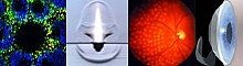
Die Arbeitsgruppe um Robert Huber forscht in den Bereichen der optischen Kohärenztomographie (OCT), der nichtlinearen Bildgebung und der Laserphysik. Hierbei wird vor allem an neuartigen Pikosekundenlaser und an Fourier domain modengekoppelten (FDML) Lasern gearbeitet. Dieses Laserkonzept wurde von Robert Huber entwickelt und erlaubt, besonders schnell durchstimmbare Laserlichtquellen zu realisieren. Die Forschungsschwerpunkte liegen hier in der technologischen Weiterentwicklung, dem Verständnis der physikalischen Vorgänge und auch in der Implementierung von FDML Lasern für OCT. Neben der OCT werden FDML Laser auch für die nichtlineare Bildgebung und Spektroskopie verwendet.
Ein weiterer Schwerpunkt liegt in der optischen Kohärenztomographie mit durchstimmbaren Lichtquellen (swept source OCT, SS-OCT). Hier werden unter anderem die selbst entwickelten FDML Laser für die ultraschnelle Bildgebung eingesetzt, um Schnittbilder von biologischen Gewebe wie Haut oder Auge zu erzeugen. Aufgrund der hohen Durchstimmrate sind Anwendungen wie die VR-OCT, die in einer virtuellen Umgebung ganze Volumina mit Video-Wiederholraten in Echtzeit darstellt, möglich.
Mit der nichtlinearen optischen Bildgebung verfolgt die Arbeitsgruppe weitere Bildgebungsverfahren. Die Forschungsgebiete liegen in der zeitcodierten (TICO) Ramanspektroskopie und -mikroskopie, der Zweiphotonen-Fluoreszenzmikroskopie (TPEF) und der Zweiphotonen-Einzelpuls-Fluoreszenzlebenszeitbildgebung (SP-FLIM). Für diese Verfahren kommen neuartige Pikosekunden zum Einsatz die ebenfalls in der Arbeitsgruppe erforscht und entwickelt werden.
Unsere Forschungsschwerpunkte:
- Fourier Domain Mode Locked (FDML) Laser - Laserphysik, Technologie und Anwendung
- Optische Kohärenztomographie (OCT) - Anwendungen der MHz-OCT an Haut und Auge
- Nichtlineare Mikroskopie und Spektroskopie
- Pikosekundenlaser
Publikationen
2023
Dual Amplification 850 nm FDML Laser, in 2023 Conference on Lasers and Electro-Optics Europe & European Quantum Electronics Conference (CLEO/Europe-EQEC) , Jul.2023. pp. 1.
| DOI: | 10.1109/CLEO/Europe-EQEC57999.2023.10232019 |
| Bibtex: |  @INPROCEEDINGS{10232019,
author={Klufts, M. and Lotz, S. and Bashir, M. A. and Pfeiffer, T. and Mlynek, A. and Wieser, W. and Chamorovskiy, A. and Shidlovski, V. and Podoleanu, A. and Huber, R.},
booktitle={2023 Conference on Lasers and Electro-Optics Europe & European Quantum Electronics Conference (CLEO/Europe-EQEC)},
title={Dual Amplification 850 nm FDML Laser},
year={2023},
volume={},
number={},
pages={1-1},
doi={10.1109/CLEO/Europe-EQEC57999.2023.10232019}}
|
Four-Wave Mixing Fast Wavelength Sweeping FDML Laser with kW Peak Power at 900 nm and 1300 nm, in 2023 Conference on Lasers and Electro-Optics Europe & European Quantum Electronics Conference (CLEO/Europe-EQEC) , Jul.2023. pp. 1.
| DOI: | 10.1109/CLEO/Europe-EQEC57999.2023.10232141 |
| Bibtex: | @INPROCEEDINGS{10232141,
author={Lamminger, Philipp and Hakert, Hubertus and Lotz, Simon and Kolb, Jan Philip and Kutscher, Tonio and Karpf, Sebastian and Huber, Robert},
booktitle={2023 Conference on Lasers and Electro-Optics Europe & European Quantum Electronics Conference (CLEO/Europe-EQEC)},
title={Four-Wave Mixing Fast Wavelength Sweeping FDML Laser with kW Peak Power at 900 nm and 1300 nm},
year={2023},
volume={},
number={},
pages={1-1},
doi={10.1109/CLEO/Europe-EQEC57999.2023.10232141}}
|
1190 nm FDML laser: Challenges and Strategies, in 2023 Conference on Lasers and Electro-Optics Europe & European Quantum Electronics Conference (CLEO/Europe-EQEC) , Jul.2023. pp. 1.
| DOI: | 10.1109/CLEO/Europe-EQEC57999.2023.10232661 |
| Bibtex: | @INPROCEEDINGS{10232661,
author={Bashir, M. A. and Lotz, S. and Kluftsa, M. and Jirauschek, C. and Huberab, R.},
booktitle={2023 Conference on Lasers and Electro-Optics Europe & European Quantum Electronics Conference (CLEO/Europe-EQEC)},
title={1190 nm FDML laser: Challenges and Strategies},
year={2023},
volume={},
number={},
pages={1-1},
doi={10.1109/CLEO/Europe-EQEC57999.2023.10232661}}
|
Four-wave mixing seeded by a rapid wavelength-sweeping FDML laser for nonlinear imaging at 900 nm and 1300 nm, Opt. Lett. , vol. 48, no. 14, pp. 3713-3716, Jul. 2023. Optica Publishing Group.
| DOI: | 10.1364/OL.488181 |
| Bibtex: | @article{Lamminger:23,
author = {Philipp Lamminger and Hubertus Hakert and Simon Lotz and Jan Philip Kolb and Tonio Kutscher and Sebastian Karpf and Robert Huber},
journal = {Opt. Lett.},
keywords = {Green fluorescent protein; Laser beam combining; Laser crystals; Laser imaging; Optical amplifiers; Photonic crystal lasers},
number = {14},
pages = {3713--3716},
publisher = {Optica Publishing Group},
title = {Four-wave mixing seeded by a rapid wavelength-sweeping FDML laser for nonlinear imaging at 900 nm and 1300 nm},
volume = {48},
month = {Jul},
year = {2023},
url = {https://opg.optica.org/ol/abstract.cfm?URI=ol-48-14-3713},
doi = {10.1364/OL.488181},
abstract = {Four-wave mixing (FWM) enables the generation and amplification of light in spectral regions where suitable fiber gain media are unavailable. The 1300 nm and 900 nm regions are of especially high interest for time-encoded (TICO) stimulated Raman scattering microscopy and spectro-temporal laser imaging by diffracted excitation (SLIDE) two-photon microscopy. We present a new, to the best of our knowledge, FWM setup where we shift the power of a home-built fully fiber-based master oscillator power amplifier (MOPA) at 1064 nm to the 1300-nm region of a rapidly wavelength-sweeping Fourier domain mode-locked (FDML) laser in a photonic crystal fiber (PCF) creating pulses in the 900-nm region. The resulting 900-nm light can be wavelength swept over 54 nm and has up to 2.5 kW (0.2 {\textmu}J) peak power and a narrow instantaneous spectral linewidth of 70 pm. The arbitrary pulse patterns of the MOPA and the fast wavelength tuning of the FDML laser (419 kHz) allow it to rapidly tune the FWM light enabling new and faster TICO-Raman microscopy, SLIDE imaging, and other applications.},
} |
The neurosurgical benefit of contactless in vivo optical coherence tomography regarding residual tumor detection: A clinical study, Frontiers in Oncology , vol. 13, Apr. 2023.
| DOI: | 10.3389/fonc.2023.1151149 |
| Datei: | fonc.2023.1151149 |
| Bibtex: | Kuppler P, Strenge P, Lange B, Spahr-Hess S, Draxinger W, Hagel C, Theisen-Kunde D, Brinkmann R, Huber R, Tronnier V and Bonsanto MM (2023) The neurosurgical benefit of contactless in vivo optical coherence tomography regarding residual tumor detection: A clinical study. Front. Oncol. 13:1151149. doi: 10.3389/fonc.2023.1151149 |
High speed 4D in-vivo OCT imaging of the human brain: creating high density datasets for machine learning toward identification of malign tissue in real time, in High-Speed Biomedical Imaging and Spectroscopy VIII , Kevin K. Tsia and Keisuke Goda, Eds. SPIE, Mä.2023. pp. 123900D.
| DOI: | 10.1117/12.2648505 |
| Bibtex: | @inproceedings{10.1117/12.2648505,
author = {Wolfgang Draxinger and Dirk Theisen-Kunde and Lion Sch{\"u}tzeck and Nicolas Detrez and Paul Strenge and Veit Danicke and Jessica Kren and Patrick Kuppler and Sonja Spahr-Hess and Matteo Mario Bonsanto and Ralf Brinkmann and Robert Huber},
title = {{High speed 4D in-vivo OCT imaging of the human brain: creating high density datasets for machine learning toward identification of malign tissue in real time}},
volume = {12390},
booktitle = {High-Speed Biomedical Imaging and Spectroscopy VIII},
editor = {Kevin K. Tsia and Keisuke Goda},
organization = {International Society for Optics and Photonics},
publisher = {SPIE},
pages = {123900D},
abstract = {Neuro-surgery is challenged by the difficulties of determining brain tumor boundaries during excisions. Optical coherence tomography is investigated as an imaging modality for providing a viable contrast channel. Our MHz-OCT technology enables rapid volumetric imaging, suitable for surgical workflows. We present a surgical microscope integrated MHz-OCT imaging system, which is used for the collection of in-vivo images of human brains, with the purpose of being used in machine learning systems that shall be trained to identify and classify tumorous tissue.},
keywords = {optical coherence tomography, brain tumor, neurosurgery, machine learning, contrast augmentation, histology dataset, clinical study, in-vivo imaging},
year = {2023},
doi = {10.1117/12.2648505},
URL = {https://doi.org/10.1117/12.2648505}
} |
Ultrafast Spectral Tuning of a Fiber Laser for Time-Encoded Multiplex Coherent Raman Scattering Microscopy, The Journal of Physical Chemistry B , pp. null, Mä. 2023.
| DOI: | 10.1021/acs.jpcb.2c09115 |
| Datei: | acs.jpcb.2c09115 |
| Bibtex: | @article{doi:10.1021/acs.jpcb.2c09115,
author = {Gottschall, Thomas and Meyer-Zedler, Tobias and Eibl, Matthias and Pfeiffer, Tom and Hakert, Hubertus and Schmitt, Michael and Huber, Robert and Tünnermann, Andreas and Limpert, Jens and Popp, Juergen},
title = {Ultrafast Spectral Tuning of a Fiber Laser for Time-Encoded Multiplex Coherent Raman Scattering Microscopy},
journal = {The Journal of Physical Chemistry B},
volume = {0},
number = {0},
pages = {null},
year = {0},
doi = {10.1021/acs.jpcb.2c09115},
note ={PMID: 36917762},
URL = {https://doi.org/10.1021/acs.jpcb.2c09115},
eprint = { https://doi.org/10.1021/acs.jpcb.2c09115}
} |
1190 nm Fourier domain mode locked (FDML) laser for optical coherence tomography (OCT), in Optical Coherence Tomography and Coherence Domain Optical Methods in Biomedicine XXVII , Joseph A. Izatt and James G. Fujimoto, Eds. SPIE, Mä.2023. pp. 1236707.
| DOI: | 10.1117/12.2652884 |
| Bibtex: | @inproceedings{10.1117/12.2652884,
author = {M. A. Bashir and S. Lotz and M. Klufts and I. Krestnikov and C. Jirauschek and R. Huber},
title = {{1190 nm Fourier domain mode locked (FDML) laser for optical coherence tomography (OCT)}},
volume = {12367},
booktitle = {Optical Coherence Tomography and Coherence Domain Optical Methods in Biomedicine XXVII},
editor = {Joseph A. Izatt and James G. Fujimoto},
organization = {International Society for Optics and Photonics},
publisher = {SPIE},
pages = {1236707},
abstract = {We demonstrate a Fourier domain mode locked (FDML) laser centered at 1190 nm with 2×410 kHz sweep repetition rate, a sweeping range of 100 nm and 2.5 mW output power. The laser is based on a quantum dot-semiconductor optical amplifier with small linewidth enhancement factor. The laser could be used as a probe laser in stimulated Raman scattering microscopy and it may be attractive for optical coherence tomography due to low water absorption and the spectral signature of lipids around 1200nm. Moreover, it is ideal to close the gap between FDML lasers at 1064 nm and 1300 nm. Combining these three lasers can enable ultrawideband sweeping to improve the axial OCT resolution down to 2 μm. },
keywords = {FDML, Swept source, laser, SS-OCT, OCT, Tunable lasers},
year = {2023},
doi = {10.1117/12.2652884},
URL = {https://doi.org/10.1117/12.2652884}
}
|
850 nm FDML: performance and challenges, in Optical Coherence Tomography and Coherence Domain Optical Methods in Biomedicine XXVII , Joseph A. Izatt and James G. Fujimoto, Eds. SPIE, Mä.2023. pp. 1236705.
| DOI: | 10.1117/12.2649646 |
| Bibtex: | @inproceedings{10.1117/12.2649646,
author = {M. Klufts and S. Lotz and M. A. Bashir and T. Pfeiffer and A. Mlynek and W. Wieser and A. Chamorovskiy and V. Shidlovski and R. Huber},
title = {{850 nm FDML: performance and challenges}},
volume = {12367},
booktitle = {Optical Coherence Tomography and Coherence Domain Optical Methods in Biomedicine XXVII},
editor = {Joseph A. Izatt and James G. Fujimoto},
organization = {International Society for Optics and Photonics},
publisher = {SPIE},
pages = {1236705},
abstract = {We demonstrate a Fourier domain mode locked (FDML) laser centered around 850 nm with a sweeping range of 50 nm, a fundamental repetition rate of 2×416 kHz and an output power of 2 mW. A new cavity design using three chirped Fiber Bragg gratings is required to overcome sweeping limitations caused by high dispersion. Other solutions to address challenges such as high loss and high polarization mode dispersion will be discussed along with performance. A main application of this laser will be retinal imaging, but it might also be applicable for TiCo-Raman and SLIDE microscopy. },
keywords = {Swept source, FDML, Laser, Ophthalmic imaging, OCT, 800 nm, retinal imaging, light sources},
year = {2023},
doi = {10.1117/12.2649646},
URL = {https://doi.org/10.1117/12.2649646}
}
|
900 nm swept source FDML laser with kW peak power, in Fiber Lasers XX: Technology and Systems , V. R. Supradeepa, Eds. SPIE, Mä.2023. pp. 124001I.
| DOI: | 10.1117/12.2649663 |
| Bibtex: | @inproceedings{10.1117/12.2649663,
author = {Philipp Lamminger and Hubertus Hakert and Simon Lotz and Jan Philip Kolb and Tonio Kutscher and Sebastian Karpf and Robert Huber},
title = {{900 nm swept source FDML laser with kW peak power}},
volume = {12400},
booktitle = {Fiber Lasers XX: Technology and Systems},
editor = {V. R. Supradeepa},
organization = {International Society for Optics and Photonics},
publisher = {SPIE},
pages = {124001I},
abstract = {A wavelength agile 900 nm 2.5 kW peak power fiber laser is created by four-wave mixing (FWM) in a photonic crystal fiber (PCF), while amplifying a 1300 nm Fourier-domain mode-locked (FDML) laser. The FWM process is pumped by a home-built 1064 nm master oscillator power amplifier (MOPA) laser and seeded by a home-built 1300 nm FDML laser, generating high power pulses at wavelengths, where amplification by active fiber media is difficult. The 900 nm pulses have a spectral linewidth of 70 pm, are tunable over 54 nm and have electronic pulse-to-pulse tuning capability. These pulses can be used for nonlinear imaging like two-photon or coherent anti-Stokes Raman microscopy (CARS) microscopy including spectro-temporal laser imaging by diffracted excitation (SLIDE) and time-encoded (Tico) stimulated Raman microscopy.},
keywords = {Fourier domain mode locking, FDML, Raman, two photon microscopy, SLIDE, 900 nm, fiber laser, photonic crystal fiber, swept source},
year = {2023},
doi = {10.1117/12.2649663},
URL = {https://doi.org/10.1117/12.2649663}
} |
Characterization of brain tumor tissue by time-resolved, phase-sensitive optical coherence elastography at 3.2 MHz line rate, in Advanced Biomedical and Clinical Diagnostic and Surgical Guidance Systems XXI , Caroline Boudoux and James W. Tunnell, Eds. SPIE, Mä.2023. pp. 123680F.
| DOI: | 10.1117/12.2648301 |
| Bibtex: | @inproceedings{10.1117/12.2648301,
author = {Sazgar Burhan and Nicolas Detrez and Katharina Rewerts and Madita G{\"o}b and Christian Hagel and Matteo Mario Bonsanto and Dirk Theisen-Kunde and Robert Huber and Ralf Brinkmann},
title = {{Characterization of brain tumor tissue by time-resolved, phase-sensitive optical coherence elastography at 3.2 MHz line rate}},
volume = {12368},
booktitle = {Advanced Biomedical and Clinical Diagnostic and Surgical Guidance Systems XXI},
editor = {Caroline Boudoux and James W. Tunnell},
organization = {International Society for Optics and Photonics},
publisher = {SPIE},
pages = {123680F},
abstract = {Optical coherence elastography (OCE) offers the possibility of obtaining the mechanical behavior of a tissue. When also using a non-contact mechanical excitation, it mimics palpation without interobserver variability. One of the most frequently used techniques is phase-sensitive OCE. Depending on the system, depth-resolved changes in the sub-µm to nm range can be detected and visualized volumetrically. Such an approach is used in this work to investigate and detect transitions between healthy and tumorous brain tissue as well as inhomogeneities in the tumor itself to assist the operating surgeon during tumor resection in the future. We present time-resolved, phase-sensitive OCE measurements on various ex vivo brain tumor samples using an ultra-fast 3.2 MHz swept-source optical coherence tomography (SS-OCT) system with a frame rate of 2.45 kHz. 4 mm line scans are acquired which, in combination with the high imaging speed, allow monitoring and investigation of the sample's behavior in response to the mechanical load. Therefore, an air-jet system applies a 200 ms short air pulse to the sample, whose non-contact property facilitates the possibility for future in vivo measurements. Since we can temporally resolve the response of the sample over the entire acquisition time, the mechanical properties are evaluated at different time points with depth resolution. This is done by unwrapping the phase data and performing subsequent assessment. Systematic ex vivo brain tumor measurements were conducted and visualized as distribution maps. The study outcomes are supported by histological analyses and examined in detail.},
keywords = { Optical Coherence Tomography, Optical Coherence Elastography, Phase-sensitive OCT, Fourier Domain Mode Locking, Brain Tumor, Phase Unwrapping, Tissue Characterization, Biomechanics},
year = {2023},
doi = {10.1117/12.2648301},
URL = {https://doi.org/10.1117/12.2648301}
}
|
Large area robotically assisted optical coherence tomography (LARA-OCT) for skin imaging with MHz-OCT surface tracking, in Optical Coherence Tomography and Coherence Domain Optical Methods in Biomedicine XXVII , Joseph A. Izatt and James G. Fujimoto, Eds. SPIE, Mä.2023. pp. 123670C.
| DOI: | 10.1117/12.2652616 |
| Bibtex: | @inproceedings{10.1117/12.2652616,
author = {Madita G{\"o}b and Simon Lotz and Linh Ha-Wissel and Sazgar Burhan and Sven B{\"o}ttger and Floris Ernst and Jennifer Hundt and Robert Huber},
title = {{Large area robotically assisted optical coherence tomography (LARA-OCT) for skin imaging with MHz-OCT surface tracking}},
volume = {12367},
booktitle = {Optical Coherence Tomography and Coherence Domain Optical Methods in Biomedicine XXVII},
editor = {Joseph A. Izatt and James G. Fujimoto},
organization = {International Society for Optics and Photonics},
publisher = {SPIE},
pages = {123670C},
abstract = {Optical coherence tomography (OCT) is a powerful imaging technique to non-invasively differentiate between healthy skin and pathological conditions. Unfortunately, commercially available OCT-systems are typically slow and not capable of scanning large areas at reasonable speed. Since skin lesions may extend over several square centimeters, potential inflammatory infiltrates remain undetected. Here, we present large area robotically assisted OCT (LARA-OCT) for skin imaging. Therefor a collaborative robot is combined with an existing, home-built 3.3 MHz-OCT-system and for surface tracking an online probe-to-surface control is implemented which is solely based on the OCT surface signal. It features a combined surface-distance and surface-orientation closed-loop control algorithm, which enables automatic positioning and alignment of the probe across the target while imaging. This allows to acquire coherent OCT images of skin areas beyond 10 cm<sup>2</sup>. },
keywords = {Optical Coherence Tomography, Fourier Domain Mode Locking, Robotically Assisted Imaging Systems, Three-dimensional image acquisition, Large Area Scanning, Skin Imaging , OCT, FDML},
year = {2023},
doi = {10.1117/12.2652616},
URL = {https://doi.org/10.1117/12.2652616}
}
|
Synchronous high-speed OCT imaging with sensor less brushless DC motor and FDML laser in a phase-locked loop, in Optical Coherence Tomography and Coherence Domain Optical Methods in Biomedicine XXVII , Joseph A. Izatt and James G. Fujimoto, Eds. SPIE, Mä.2023. pp. 1236703.
| DOI: | 10.1117/12.2652955 |
| Bibtex: | @inproceedings{10.1117/12.2652955,
author = {Awanish Pratap Singh and Madita G{\"o}b and Martin Ahrens and Tim Eixmann and Hinnerk Schulz-Hildebrandt and Gereon H{\"u}ttmann and Robert Huber and Maik Rahlves},
title = {{Synchronous high-speed OCT imaging with sensor less brushless DC motor and FDML laser in a phase-locked loop}},
volume = {12367},
booktitle = {Optical Coherence Tomography and Coherence Domain Optical Methods in Biomedicine XXVII},
editor = {Joseph A. Izatt and James G. Fujimoto},
organization = {International Society for Optics and Photonics},
publisher = {SPIE},
pages = {1236703},
abstract = {High-speed endoscopic optical coherence tomography (OCT) imaging in the MHz range has shown great potential in various medical applications ranging from cancer screening to vascular disease monitoring. High-speed imaging always suffers from non-uniform rotational distortion (NURD) due to asynchronous motor rotation with the OCT system. Several research groups have previously attempted to solve this problem, using either an expensive motor with a sensor or numerical correction after data acquisition. However, both techniques pose challenges for practical use. Therefore, in this study, we use an inexpensive sensorless brushless DC motor with a Fourier domain mode-locked (FDML) laser-based MHz OCT system and try to resolve the problem of synchronization using three different modalities, (i) Slave-mode: The FDML frequency serves as a master frequency for the motor, which is phase-locked to the FDML frequency, (ii) Master-mode: The revolution trigger obtained from the motor’s back electromotive force (BEMF) signal serves as a trigger signal for the OCT imaging system, (iii) Both: Fully synchronized setup, where the motor rotation is synchronized with the laser and the imaging system is synchronized with the motor to achieve phase-stable OCT imaging. The first case slightly fluctuates in live preview and imaging due to the absence of a revolution trigger, while the second has varying motor speeds. Therefore, we use the third case to phase-lock the motor with FDML and get a distortion-free live preview and image acquisition. Finally, we demonstrate high-speed SS-OCT structural imaging (at 3.3 MHz A-scan rates) of a finger with a 16 mm diameter probe (at 40,000 rpm).},
keywords = {Optical Coherence Tomography, Endoscopy, FDML , Closed Loop Motor Control, NURD compensation, Brushless DC Motor, Back Electromotive Force},
year = {2023},
doi = {10.1117/12.2652955},
URL = {https://doi.org/10.1117/12.2652955}
} |
Dual wavelength analysis and classification of brain tumor tissue with optical coherence tomography, in Advanced Biomedical and Clinical Diagnostic and Surgical Guidance Systems XXI , Caroline Boudoux and James W. Tunnell, Eds. SPIE, Mä.2023. pp. 1236805.
| DOI: | 10.1117/12.2649963 |
| Bibtex: | @inproceedings{10.1117/12.2649963,
author = {Paul Strenge and Birgit Lange and Wolfgang Draxinger and Christian Hagel and Christin Grill and Veit Danicke and Dirk Theisen-Kunde and Sonja Spahr-Hess and Matteo M. Bonsanto and Robert Huber and Heinz Handels and Ralf Brinkmann},
title = {{Dual wavelength analysis and classification of brain tumor tissue with optical coherence tomography}},
volume = {12368},
booktitle = {Advanced Biomedical and Clinical Diagnostic and Surgical Guidance Systems XXI},
editor = {Caroline Boudoux and James W. Tunnell},
organization = {International Society for Optics and Photonics},
publisher = {SPIE},
pages = {1236805},
abstract = {The ill-defined tumor borders of glioblastoma multiforme pose a major challenge for the surgeon during tumor resection, since the goal of the tumor resection is the complete removal, while saving as much healthy brain tissue as possible. In recent years, optical coherence tomography (OCT) was successfully used to classify white matter from tumor infiltrated white matter by several research groups. Motivated by these results, a dataset was created, which consisted of sets of corresponding ex vivo OCT images, which were acquired by two OCT-systems with different properties (e.g. wavelength and resolution). Each image was annotated with semantic labels. The labels differentiate between white and gray matter and three different stages of tumor infiltration. The data from both systems not only allowed a comparison of the ability of a system to identify the different tissue types present during the tumor resection, but also enable a multimodal tissue analysis evaluating corresponding OCT images of the two systems simultaneously. A convolutional neural network with dirichlet prior was trained, which allowed to capture the uncertainty of a prediction. The approach increased the sensitivity of identifying tumor infiltration from 58 % to 78 % for data with a low prediction uncertainty compared to a previous monomodal approach. },
keywords = {optical coherence tomography, oct, brain, classification, tumor, dual wavelength, glioblastoma multiforme, tissue analysis},
year = {2023},
doi = {10.1117/12.2649963},
URL = {https://doi.org/10.1117/12.2649963}
} |
Accelerating intraoperative tumor histology with sectioning-free multiphoton microscopy, European Journal of Surgical Oncology , vol. 49, no. 2, pp. e210, Feb. 2023.
| DOI: | https://doi.org/10.1016/j.ejso.2022.11.575 |
| Datei: | S0748798322013245 |
| Bibtex: | @article{STRAUCH2023e210,
title = {Accelerating intraoperative tumor histology with sectioning-free multiphoton microscopy},
journal = {European Journal of Surgical Oncology},
volume = {49},
number = {2},
pages = {e210},
year = {2023},
issn = {0748-7983},
doi = {https://doi.org/10.1016/j.ejso.2022.11.575},
url = {https://www.sciencedirect.com/science/article/pii/S0748798322013245},
author = {Matthias Strauch and Jan Philip Kolb and Christian Rose and Nadine Merg and Jennifer Hundt and Christiane Kümpers and Sven Perner and Sebastian Karpf and Robert Huber}
} |
Air-jet based optical coherence elastography of brain tumor tissue: stiffness evaluation by structural histological analysis, in Emerging Technologies for Cell and Tissue Characterization II , Seemantini K. Nadkarni and Giuliano Scarcelli, Eds. SPIE, 2023. pp. 126290M.
| DOI: | 10.1117/12.2670944 |
| Datei: | 12.2670944 |
| Bibtex: | @inproceedings{10.1117/12.2670944,
author = {Nicolas Detrez and Sazgar Burhan and Paul Strenge and Jessica Kren and Christian Hagel and Matteo Mario Bonsanto and Dirk Theisen-Kunde and Robert Huber and Ralf Brinkmann},
title = {{Air-jet based optical coherence elastography of brain tumor tissue: stiffness evaluation by structural histological analysis}},
volume = {12629},
booktitle = {Emerging Technologies for Cell and Tissue Characterization II},
editor = {Seemantini K. Nadkarni and Giuliano Scarcelli},
organization = {International Society for Optics and Photonics},
publisher = {SPIE},
pages = {126290M},
keywords = {Optical Coherence Elastography, Air-Jet, Phase-sensitive OCT, Histology Structure Analysis, Color-Deconvolution, Structural Tensors, Brain tumor, Tissue Characterization},
year = {2023},
doi = {10.1117/12.2670944},
URL = {https://doi.org/10.1117/12.2670944}
} |
Air-Jet based optical coherence elastography: processing and mechanical interpretation of brain tumor data, in Optical Elastography and Tissue Biomechanics X , Kirill V. Larin and Giuliano Scarcelli and Frédérique Vanholsbeeck, Eds. SPIE, 2023. pp. 1238105.
| DOI: | 10.1117/12.2649835 |
| Datei: | 12.2649835 |
| Bibtex: | @inproceedings{10.1117/12.2649835,
author = {Nicolas Detrez and Sazgar Burhan and Katharina Rewerts and Jessica Kren and Christian Hagel and Matteo Mario Bonsanto and Dirk Theisen-Kunde and Robert Huber and Ralf Brinkmann},
title = {{Air-Jet based optical coherence elastography: processing and mechanical interpretation of brain tumor data}},
volume = {12381},
booktitle = {Optical Elastography and Tissue Biomechanics X},
editor = {Kirill V. Larin and Giuliano Scarcelli and Fr{\'e}d{\'e}rique Vanholsbeeck},
organization = {International Society for Optics and Photonics},
publisher = {SPIE},
pages = {1238105},
keywords = {Optical Coherence Elastography, Air-Jet, Air-Puff, biomechanics, viscoelasticity, rheology, brain tissue, brain tumor},
year = {2023},
doi = {10.1117/12.2649835},
URL = {https://doi.org/10.1117/12.2649835}
} |
MHz time stretch swept source using a commercial erbium-doped fiber amplifier, in Optical Coherence Tomography and Coherence Domain Optical Methods in Biomedicine XXVII , Joseph A. Izatt and James G. Fujimoto, Eds. SPIE, 2023. pp. 1236706.
| DOI: | 10.1117/12.2651127 |
| Datei: | 12.2651127 |
| Bibtex: | @inproceedings{10.1117/12.2651127,
author = {A. Mart{\'i}nez Jim{\'e}nez and M. Spacek and M. Wacker and R. Huber and A. Bradu and A. Podoleanu},
title = {{MHz time stretch swept source using a commercial erbium-doped fiber amplifier}},
volume = {12367},
booktitle = {Optical Coherence Tomography and Coherence Domain Optical Methods in Biomedicine XXVII},
editor = {Joseph A. Izatt and James G. Fujimoto},
organization = {International Society for Optics and Photonics},
publisher = {SPIE},
pages = {1236706},
keywords = {swept source, time-stretch, optical coherence tomography, mode-locking},
year = {2023},
doi = {10.1117/12.2651127},
URL = {https://doi.org/10.1117/12.2651127}
} |
Phase analysis strategies for MHz OCE in the large displacement regime, in Optical Coherence Tomography and Coherence Domain Optical Methods in Biomedicine XXVII , Joseph A. Izatt and James G. Fujimoto, Eds. SPIE, 2023. pp. 123670Q.
| DOI: | 10.1117/12.2652847 |
| Bibtex: | @inproceedings{10.1117/12.2652847,
author = {Sazgar Burhan and Nicolas Detrez and Katharina Rewerts and Madita G{\"o}b and Steffen Buschschl{\"u}ter and Christian Hagel and Matteo Mario Bonsanto M.D. and Dirk Theisen-Kunde and Robert Huber and Ralf Brinkmann},
title = {{Phase analysis strategies for MHz OCE in the large displacement regime}},
volume = {12367},
booktitle = {Optical Coherence Tomography and Coherence Domain Optical Methods in Biomedicine XXVII},
editor = {Joseph A. Izatt and James G. Fujimoto},
organization = {International Society for Optics and Photonics},
publisher = {SPIE},
pages = {123670Q},
abstract = {In neurosurgical tumor operations on the central nervous system, intraoperative haptic information often assists for discrimination between healthy and diseased tissue. Thus, it can provide the neurosurgeon with additional intraoperative source of information during resection, next to the visual information by the light microscope, fluorescent dyes and neuronavigation. One approach to obtain elastic and viscoelastic tissue characteristics non-subjectively is phase-sensitive optical coherence elastography (OCE), which is based on the principle of optical coherence tomography (OCT). While phase-sensitive OCE offers significantly higher displacement sensitivity inside a sample than commonly used intensity-based correlation methods, it requires a reliable algorithm to recover the phase signal, which is mathematically restricted in the -π to π range. This problem of phase wrapping is especially critical for inter-frame phase analysis since the time intervals between two referenced voxels is long. Here, we demonstrate a one-dimensional unwrapping algorithm capable of removing up to 4π-ambiguities between two frames in the complex phase data obtained from a 3.2 MHz-OCT system. The high sampling rate allows us to resolve large sample displacements induced by a 200 ms air pulse and acquires pixel-precise detail information. The deformation behavior of the tissue can be monitored over the entire acquisition time, offering various subsequent mechanical analysis procedures. The reliability of the algorithm and imaging concept was initially evaluated using different brain tumor mimicking phantoms. Additionally, results from human ex vivo brain tumor samples are presented and correlated with histological findings supporting the robustness of the algorithm.},
keywords = {Optical Coherence Tomography, Megahertz OCT, Fourier Domain Mode Locking, Optical Coherence Elastography, Phase-sensitive OCT, Phase Unwrapping, Brain tumor, Biomechanics},
year = {2023},
doi = {10.1117/12.2652847},
URL = {https://doi.org/10.1117/12.2652847}
} |
2022
Influence of the linewidth enhancement factor on the signal pattern of Fourier domain mode-locked lasers, Applied Physics B , vol. 18(12), Nov. 2022.
| DOI: | 10.1007/s00340-022-07933-5 |
| Bibtex: | @article{RN5426,
author = {Aşırım, Özüm Emre;Huber, Robert and Jirauschek, Christian},
title = {Influence of the linewidth enhancement factor on the signal pattern of Fourier domain mode-locked lasers},
journal = {Applied Physics B},
volume = {128},
number = {12},
pages = {218},
ISSN = {1432-0649},
DOI = {10.1007/s00340-022-07933-5},
url = {https://doi.org/10.1007/s00340-022-07933-5},
year = {2022},
type = {Journal Article}
}
|
620 Screening an inhibitor library for new drug candidates to promote wound healing, Journal of Investigative Dermatology , vol. 142, no. 12, Supplement, pp. S288, Nov. 2022.
| DOI: | https://doi.org/10.1016/j.jid.2022.09.637 |
| Datei: | S0022202X22025714 |
| Bibtex: | @article{JACOBI2022S288,
title = {620 Screening an inhibitor library for new drug candidates to promote wound healing},
journal = {Journal of Investigative Dermatology},
volume = {142},
number = {12, Supplement },
pages = {S288},
year = {2022},
note = {ESDR 2022 Meeting Abstract Supplement},
issn = {0022-202X},
doi = {https://doi.org/10.1016/j.jid.2022.09.637},
url = {https://www.sciencedirect.com/science/article/pii/S0022202X22025714},
author = {C. Jacobi and M. Göb and R. Huber and R.J. Ludwig and J.E. Hundt}
} |
Intravascular optical coherence elastography, Biomed. Opt. Express , vol. 13, no. 10, pp. 5418--5433, Okt. 2022. Optica Publishing Group.
| DOI: | 10.1364/BOE.470039 |
| Datei: | abstract.cfm |
| Bibtex: | Optical coherence elastography (OCE), a functional extension of optical coherence tomography (OCT), visualizes tissue strain to deduce the tissue’s biomechanical properties. In this study, we demonstrate intravascular OCE using a 1.1 mm motorized catheter and a 1.6 MHz Fourier domain mode-locked OCT system. We induced an intraluminal pressure change by varying the infusion rate from the proximal end of the catheter. We analysed the pixel-matched phase change between two different frames to yield the radial strain. Imaging experiments were carried out in a phantom and in human coronary arteries in vitro. At an imaging speed of 3019 frames/s, we were able to capture the dynamic strain. Stiff inclusions in the phantom and calcification in atherosclerotic plaques are associated with low strain values and can be distinguished from the surrounding soft material, which exhibits elevated strain. For the first time, circumferential intravascular OCE images are provided side by side with conventional OCT images, simultaneously mapping both the tissue structure and stiffness. |
Case report: Optical coherence tomography for monitoring biologic therapy in psoriasis and atopic dermatitis, Frontiers in Medicine , vol. 9, Sep. 2022.
| DOI: | 10.3389/fmed.2022.995883 |
| Datei: | fmed.2022.995883 |
| Bibtex: | @article{RN5359,
author = {Ha-Wissel, L.;Yasak, H.;Huber, R.;Zillikens, D.;Ludwig, R. J.;Thaçi, D. and Hundt, J. E.},
title = {Case report: Optical coherence tomography for monitoring biologic therapy in psoriasis and atopic dermatitis},
journal = {Front Med (Lausanne)},
volume = {9},
pages = {995883},
ISSN = {2296-858X (Print)
2296-858x},
DOI = {10.3389/fmed.2022.995883},
year = {2022},
type = {Journal Article}
}
|
Differentiation of different stages of brain tumor infiltration using optical coherence tomography: Comparison of two systems and histology, Frontiers in Oncology , Aug. 2022.
| DOI: | 10.3389/fonc.2022.896060 |
| Bibtex: | @article{Strenge-2022,
author = {Strenge, P.;Lange, B.;Grill,C.;Danicke,V.;Theisen-Kunde, D.;Hagel, C.;Spahr-Hess, S.;;Bonsanto, Matteo M.;Handels, H.; and Huber, R.;Brinkmann, R.},
title = {Differentiation of different stages of brain tumor infiltration using optical coherence tomography: Comparison of two systems and histology},
journal = {Frontiers in Oncology},
Keywords = {AG-Huber_FDML, AG-Huber_OCT, brain, tumor, glioblastoma multiforme, OCT, neural network, attenuation (absorption)
coefficient, optical coherence tomography},
DOI = {https://doi.org/10.3389/fonc.2022.896060},
url = {https://www.frontiersin.org/articles/10.3389/fonc.2022.896060/full},
year = {2022},
type = {Journal Article}
}
|
Towards phase-stabilized Fourier domain mode-locked frequency combs, Communications Physics , vol. 5, no. 1, Aug. 2022. Springer Science and Business Media LLC.
| DOI: | 10.1038/s42005-022-00960-w |
| Bibtex: | @article{Grill2022,
doi = {10.1038/s42005-022-00960-w},
year = {2022},
publisher = {Springer Science and Business Media {LLC}},
volume = {{5}},
number = {{1}},
author = {C. Grill, T. Bl\"{o}mker, M. Schmidt, D. Kastner, T. Pfeiffer, J.P. Kolb, W. Draxinger, S. Karpf, C. Jirauschek and R. Huber},
title = {Towards phase-stabilized Fourier domain mode-locked frequency combs},
journal = {{Communications Physics}},
keywords={AG-Huber_FDML, FDML, Fourier domain mode locking, phase, frequency comb, coherence, beating}
} |
Text