OCT is a noninvasive Imaging modality which is typically used for high resolution (~10µm), three dimensional imaging of scattering tissue. By using home built FDML laser technology we achieve imaging speeds of several million depth scans per second, which is one to two orders of magnitude higher than current commercially available systems (MHz-OCT).
These high imaging speeds already proved to be very useful in clinical applications, by reducing acquisition times and therefore reducing motion artifacts. But the high speed also gives access to the phase of the detected light and will thus allow the use of new numerical approaches for image quality enhancement and functional imaging with Swept-Source-OCT.
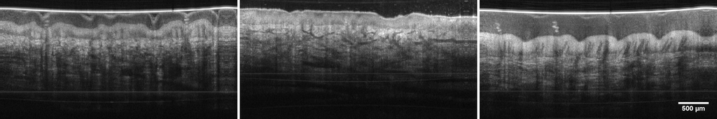
Our working group is conducting research in the field of OCT to develop new technologies and to identify possible fields of application.
The focus areas are:
- MHz-OCT - Ultra-fast OCT imaging with several million depth scans per second
- LARA-OCT - Large area robotically assisted OCT
- VR-OCT - Real-time computation and rendering of entire OCT volumes in a virtual environment
- Eye OCT - application of MHz-OCT to the eye for visualization of the retina or the anterior segment of the eye
- Phase sensitive OCT - enhancement of the information content of an OCT image by adding phase contrast
- Multispectral OCT - combination of RGB and OCT images for improved visualization of morphological structures
related Publications
2013
History compounding: a novel speckle reduction technique for OCT guided cochleostomy, in Optical Coherence Tomography and Coherence Domain Optical Methods in Biomedicine XVII , James G. Fujimoto and Joseph A. Izatt and Valery V. Tuchin, Eds. SPIE, 032013. pp. 85713H.
| DOI: | 10.1117/12.2006979 |
| Bibtex: | @inproceedings{10.1117/12.2006979,
author = {Yaokun Zhang and Tom Pfeiffer and Wolfgang Wieser and Marcel Weller and Robert Huber and Thomas Klenzner and J{\"o}rg Raczkowsky and Heinz W{\"o}rn},
title = {{History compounding: a novel speckle reduction technique for OCT guided cochleostomy}},
volume = {8571},
booktitle = {Optical Coherence Tomography and Coherence Domain Optical Methods in Biomedicine XVII},
editor = {James G. Fujimoto and Joseph A. Izatt and Valery V. Tuchin},
organization = {International Society for Optics and Photonics},
publisher = {SPIE},
pages = {85713H},
abstract = {Optical coherence tomography (OCT) is a promising candidate for monitoring the bottom of the drilled channel during
cochleostomy to prevent injury to the critical structure under the bone tissue. While the thickness of the overlaying bone
tissue is changed during the drilling process, the wave front of the backscattered light is also altered, resulting in
changing speckle patterns of the observed structures in the sequential historical scans. By averaging the different patterns
in these scans, named history compounding, the speckles can be reduced and the detection of critical structure becomes
much easier. Before averaging, the refractive index of bone tissue ???????? has to be compensated so that the speckles of the
same structure in different historical scans can be aligned together. An accurate method for measuring the refractive
index n<sub>b</sub> using OCT is presented. Experiments were conducted to evaluate history compounding and the new technique
is proved to be an effective, flexible and intuitive speckle reduction technique for OCT guided cochleostomy as well as
hard tissue ablation of other kind.},
keywords = {optical coherence tomography, speckle reduction, refractive index, cochleostomy, hard tissue ablation},
year = {2013},
doi = {10.1117/12.2006979},
URL = {https://doi.org/10.1117/12.2006979}
}
|
Retinal polarization-sensitive optical coherence tomography at 1060 nm with 350 kHz A-scan rate using an Fourier domain mode locked laser, Journal of Biomedical Optics , vol. 18, no. 2, pp. 026008, 02 2013. SPIE.
| DOI: | 10.1117/1.JBO.18.2.026008 |
| Bibtex: | @article{10.1117/1.JBO.18.2.026008,
author = {Teresa Torzicky and Sebastian Marschall and Michael Pircher and Bernhard Baumann and Marco Bonesi and Stefan Zotter and Erich G{\"o}tzinger and Wolfgang Trasischker and Thomas Klein and Wolfgang Wieser and Benjamin R. Biedermann and Robert A. Huber and Peter E. Andersen and Christoph K. Hitzenberger},
title = {{Retinal polarization-sensitive optical coherence tomography at 1060 nm with 350 kHz A-scan rate using an Fourier domain mode locked laser}},
volume = {18},
journal = {Journal of Biomedical Optics},
number = {2},
publisher = {SPIE},
pages = {026008},
abstract = {We present a novel, high-speed, polarization-sensitive, optical coherence tomography set-up for retinal imaging operating at a central wavelength of 1060 nm which was tested for in vivo imaging in healthy human volunteers. We use the system in combination with a Fourier domain mode locked laser with active spectral shaping which enables the use of forward and backward sweep in order to double the imaging speed without a buffering stage. With this approach and with a custom designed data acquisition system, we show polarization-sensitive imaging with an A-scan rate of 350 kHz. The acquired three-dimensional data sets of healthy human volunteers show different polarization characteristics in the eye, such as depolarization in the retinal pigment epithelium and birefringence in retinal nerve fiber layer and sclera. The increased speed allows imaging of large volumes with reduced motion artifacts. Moreover, averaging several two-dimensional frames allows the generation of high-definition B-scans without the use of an eye-tracking system. The increased penetration depth of the system, which is caused by the longer probing beam wavelength, is beneficial for imaging choroidal and scleral structures and allows automated segmentation of these layers based on their polarization characteristics.},
keywords = {Optical coherence tomography, Polarization, Birefringence, Imaging systems, Data acquisition, Image segmentation, Modulation, Mode locking, 3D acquisition, Retinal scanning},
year = {2013},
doi = {10.1117/1.JBO.18.2.026008},
URL = {https://doi.org/10.1117/1.JBO.18.2.026008}
}
|
Multi-MHz retinal OCT, Biomed. Opt. Express , vol. 4, no. 10, pp. 1890-1908, 2013. Optica Publishing Group.
| DOI: | 10.1364/BOE.4.001890 |
| Bibtex: | @article{Klein:13,
author = {Thomas Klein and Wolfgang Wieser and Lukas Reznicek and Aljoscha Neubauer and Anselm Kampik and Robert Huber},
journal = {Biomed. Opt. Express},
keywords = {Medical optics instrumentation; Lasers, fiber; Medical and biological imaging; Ophthalmic optics and devices ; Optical coherence tomography; Adaptive optics; Distributed Bragg reflectors; Fiber Bragg gratings; Functional imaging; Image quality; Three dimensional imaging},
number = {10},
pages = {1890--1908},
publisher = {Optica Publishing Group},
title = {Multi-MHz retinal OCT},
volume = {4},
month = {Oct},
year = {2013},
url = {https://opg.optica.org/boe/abstract.cfm?URI=boe-4-10-1890},
doi = {10.1364/BOE.4.001890},
abstract = {We analyze the benefits and problems of in vivo optical coherence tomography (OCT) imaging of the human retina at A-scan rates in excess of 1 MHz, using a 1050 nm Fourier-domain mode-locked (FDML) laser. Different scanning strategies enabled by MHz OCT line rates are investigated, and a simple multi-volume data processing approach is presented. In-vivo OCT of the human ocular fundus is performed at different axial scan rates of up to 6.7 MHz. High quality non-mydriatic retinal imaging over an ultra-wide field is achieved by a combination of several key improvements compared to previous setups. For the FDML laser, long coherence lengths and 72 nm wavelength tuning range are achieved using a chirped fiber Bragg grating in a laser cavity at 419.1 kHz fundamental tuning rate. Very large data sets can be acquired with sustained data transfer from the data acquisition card to host computer memory, enabling high-quality averaging of many frames and of multiple aligned data sets. Three imaging modes are investigated: Alignment and averaging of 24 data sets at 1.68 MHz axial line rate, ultra-dense transverse sampling at 3.35 MHz line rate, and dual-beam imaging with two laser spots on the retina at an effective line rate of 6.7 MHz.},
} |
2012
Intrasweep phase-sensitive optical coherence tomography for noncontact optical photoacoustic imaging, Opt. Lett. , vol. 37, no. 21, pp. 4368-4370, Nov. 2012. Optica Publishing Group.
| DOI: | 10.1364/OL.37.004368 |
| Bibtex: | @article{Blatter:12,
author = {Cedric Blatter and Branislav Grajciar and Pu Zou and Wolfgang Wieser and Aart-Jan Verhoef and Robert Huber and Rainer A. Leitgeb},
journal = {Opt. Lett.},
keywords = {Optical coherence tomography; Optical coherence tomography; Photoacoustic imaging; Interferometric imaging ; Photoacoustics ; In vivo imaging; Interferometry; Linewidth; Medical imaging; Optical coherence tomography; Swept sources},
number = {21},
pages = {4368--4370},
publisher = {Optica Publishing Group},
title = {Intrasweep phase-sensitive optical coherence tomography for noncontact optical photoacoustic imaging},
volume = {37},
month = {Nov},
year = {2012},
url = {https://opg.optica.org/ol/abstract.cfm?URI=ol-37-21-4368},
doi = {10.1364/OL.37.004368},
abstract = {We introduce a method to extract the photoacoustic (PA) signal from the phase time evolution of an optical coherence tomography (OCT) swept source spectral sweep. This all-optical detection is achieved in a noncontact fashion directly on the sample surface by using its specular reflection. High-speed measurement and referencing allow for close to shot noise limited phase-sensitive detection. It offers a simple way to perform OCT and PA imaging by sharing the same system components.},
} |
High-speed polarization sensitive optical coherence tomography scan engine based on Fourier domain mode locked laser, Biomed. Opt. Express , vol. 3, no. 11, pp. 2987-3000, Nov. 2012. Optica Publishing Group.
| DOI: | 10.1364/BOE.3.002987 |
| Bibtex: | @article{Bonesi:12,
author = {Marco Bonesi and Harald Sattmann and Teresa Torzicky and Stefan Zotter and Bernhard Baumann and Michael Pircher and Erich G\"{o}tzinger and Christoph Eigenwillig and Wolfgang Wieser and Robert Huber and Christoph K. Hitzenberger},
journal = {Biomed. Opt. Express},
keywords = {Optical coherence tomography; Optical diagnostics for medicine; Polarization-selective devices; High speed imaging; Image quality; Laser modes; Mode locking; Single mode fibers; Three dimensional imaging},
number = {11},
pages = {2987--3000},
publisher = {Optica Publishing Group},
title = {High-speed polarization sensitive optical coherence tomography scan engine based on Fourier domain mode locked laser},
volume = {3},
month = {Nov},
year = {2012},
url = {https://opg.optica.org/boe/abstract.cfm?URI=boe-3-11-2987},
doi = {10.1364/BOE.3.002987},
abstract = {We report on a new swept source polarization sensitive optical coherence tomography scan engine that is based on polarization maintaining (PM) fiber technology. The light source is a Fourier domain mode locked laser with a PM cavity that operates in the 1300 nm wavelength regime. It is equipped with a PM buffer stage that doubles the fundamental sweep frequency of 54.5 kHz. The fiberization allows coupling of the scan engine to different delivery probes. In a first demonstration, we use the system for imaging human skin at an A-scan rate of 109 kHz. The system illuminates the sample with circularly polarized light and measures reflectivity, retardation, optic axis orientation, and Stokes vectors simultaneously. Furthermore, depolarization can be quantified by calculating the degree of polarization uniformity (DOPU). The high scanning speed of the system enables dense sampling in both, the x- and y-direction, which provides the opportunity to use 3D evaluation windows for DOPU calculation. This improves the spatial resolution of DOPU images considerably.},
} |
Extended coherence length megahertz FDML and its application for anterior segment imaging, Biomed. Opt. Express , vol. 3, no. 10, pp. 2647-2657, Oct. 2012. Optica Publishing Group.
| DOI: | 10.1364/BOE.3.002647 |
| Bibtex: | @article{Wieser:12,
author = {Wolfgang Wieser and Thomas Klein and Desmond C. Adler and Francois Tr\'{e}panier and Christoph M. Eigenwillig and Sebastian Karpf and Joseph M. Schmitt and Robert Huber},
journal = {Biomed. Opt. Express},
keywords = {Optical coherence tomography; Lasers, tunable; Optical coherence tomography; Amplified spontaneous emission; Crystalline lens; Gastrointestinal imaging; High speed imaging; Image quality; Three dimensional imaging},
number = {10},
pages = {2647--2657},
publisher = {Optica Publishing Group},
title = {Extended coherence length megahertz FDML and its application for anterior segment imaging},
volume = {3},
month = {Oct},
year = {2012},
url = {https://opg.optica.org/boe/abstract.cfm?URI=boe-3-10-2647},
doi = {10.1364/BOE.3.002647},
abstract = {We present a 1300 nm Fourier domain mode locked (FDML) laser for optical coherence tomography (OCT) that combines both, a high 1.6 MHz wavelength sweep rate and an ultra-long instantaneous coherence length for rapid volumetric deep field imaging. By reducing the dispersion in the fiber delay line of the FDML laser, the instantaneous coherence length and hence the available imaging range is approximately quadrupled compared to previously published MHz-FDML setups, the imaging speed is increased by a factor of 16 compared to previous extended coherence length results. We present a detailed characterization of the FDML laser performance. We demonstrate for the first time MHz-OCT imaging of the anterior segment of the human eye. The OCT system provides enough imaging depth to cover the whole range from the top surface of the cornea down to the crystalline lens.},
} |
In situ structural and microangiographic assessment of human skin lesions with high-speed OCT, Biomed. Opt. Express , vol. 3, no. 10, pp. 2636-2646, Oct. 2012. Optica Publishing Group.
| DOI: | 10.1364/BOE.3.002636 |
| Bibtex: | @article{Blatter:12,
author = {Cedric Blatter and Jessika Weingast and Aneesh Alex and Branislav Grajciar and Wolfgang Wieser and Wolfgang Drexler and Robert Huber and Rainer A. Leitgeb},
journal = {Biomed. Opt. Express},
keywords = {Optical coherence tomography; Optical coherence tomography; Flow diagnostics; Functional monitoring and imaging ; Fourier domain mode locking; High speed imaging; Image processing; In vivo imaging; Speckle imaging; Three dimensional imaging},
number = {10},
pages = {2636--2646},
publisher = {Optica Publishing Group},
title = {In situ structural and microangiographic assessment of human skin lesions with high-speed OCT},
volume = {3},
month = {Oct},
year = {2012},
url = {https://opg.optica.org/boe/abstract.cfm?URI=boe-3-10-2636},
doi = {10.1364/BOE.3.002636},
abstract = {We demonstrate noninvasive structural and microvascular contrast imaging of different human skin diseases in vivo using an intensity difference analysis of OCT tomograms. The high-speed swept source OCT system operates at 1310 nm with 220 kHz A-scan rate. It provides an extended focus by employing a Bessel beam. The studied lesions were two cases of dermatitis and two cases of basal cell carcinoma. The lesions show characteristic vascular patterns that are significantly different from healthy skin. In case of inflammation, vessels are dilated and perfusion is increased. In case of basal cell carcinoma, the angiogram shows a denser network of unorganized vessels with large vessels close to the skin surface. Those results indicate that assessing vascular changes yields complementary information with important insight into the metabolic demand.},
} |
Ultrahigh-speed non-invasive widefield angiography, Journal of Biomedical Optics , vol. 17, no. 7, pp. 070505, 06 2012. SPIE.
| DOI: | 10.1117/1.JBO.17.7.070505 |
| Bibtex: | @article{10.1117/1.JBO.17.7.070505,
author = {Cedric Blatter and Branislav Grajciar and Tilman Schmoll and Rainer A. Leitgeb and Thomas Klein and Wolfgang Wieser and Raphael J. Andr{\'e} and Robert Huber},
title = {{Ultrahigh-speed non-invasive widefield angiography}},
volume = {17},
journal = {Journal of Biomedical Optics},
number = {7},
publisher = {SPIE},
pages = {070505},
abstract = {Retinal and choroidal vascular imaging is an important diagnostic benefit for ocular diseases such as age-related macular degeneration. The current gold standard for vessel visualization is fluorescence angiography. We present a potential non-invasive alternative to image blood vessels based on functional Fourier domain optical coherence tomography (OCT). For OCT to compete with the field of view and resolution of angiography while maintaining motion artifacts to a minimum, ultrahigh-speed imaging has to be introduced. We employ Fourier domain mode locking swept source technology that offers high quality imaging at an A-scan rate of up to 1.68 MHz. We present retinal angiogram over ∼ 48 deg acquired in a few seconds in a single recording without the need of image stitching. OCT at 1060 nm allows for high penetration in the choroid and efficient separate characterization of the retinal and choroidal vascularization.},
keywords = {Angiography, Optical coherence tomography, Image segmentation, Retina, Capillaries, Tissues, Visualization, Diagnostics, Gold, Vascular imaging},
year = {2012},
doi = {10.1117/1.JBO.17.7.070505},
URL = {https://doi.org/10.1117/1.JBO.17.7.070505}
}
|
High-speed polarization-sensitive OCT at 1060 nm using a Fourier domain mode-locked swept source, in Biophotonics: Photonic Solutions for Better Health Care III , Jürgen Popp and Wolfgang Drexler and Valery V. Tuchin and Dennis L. Matthews, Eds. SPIE, 052012. pp. 84271D.
| DOI: | 10.1117/12.922313 |
| Bibtex: | @inproceedings{10.1117/12.922313,
author = {Sebastian Marschall and Teresa Torzicky and Thomas Klein and Wolfgang Wieser and Michael Pircher and Erich G{\"o}tzinger and Stefan Zotter and Marco Bonesi and Benjamin Biedermann and Christian Pedersen and Robert Huber and Christoph Hitzenberger and Peter Andersen},
title = {{High-speed polarization-sensitive OCT at 1060 nm using a Fourier domain mode-locked swept source}},
volume = {8427},
booktitle = {Biophotonics: Photonic Solutions for Better Health Care III},
editor = {J{\"u}rgen Popp and Wolfgang Drexler and Valery V. Tuchin and Dennis L. Matthews},
organization = {International Society for Optics and Photonics},
publisher = {SPIE},
pages = {84271D},
abstract = {Optical coherence tomography (OCT) in the 1060nm range is interesting for in vivo imaging of the human
posterior eye segment (retina, choroid, sclera), as it permits a long penetration depth. Complementary to
structural images, polarization-sensitive OCT (PS-OCT) images visualize birefringent, polarization-maintaining
or depolarizing areas within the sample. This information can be used to distinguish retinal layers and structures
with different polarization properties. High imaging speed is crucial for imaging ocular structures in vivo in order
to minimize motion artifacts while acquiring sufficiently large datasets. Here, we demonstrate PS-OCT imaging
at 350 kHz A-scan rate using a two-channel PS-OCT system in conjunction with a Fourier domain mode-locked
laser. The light source spectrum spans up to 100nm around the water absorption minimum at 1060 nm. By
modulating the laser pump current, we can optimize the spectrum and achieve a depth resolution of 9 μm in air
(6.5 μm in tissue). We acquired retinal images in vivo with high resolution and deep penetration into choroid and
sclera, and features like the depolarizing RPE or an increasing phase retardation at the chorio-scleral interface
are clearly visualized.},
keywords = {optical coherence tomography, polarization-sensitive OCT, swept source, Fourier domain mode-locking, 1060 nm},
year = {2012},
doi = {10.1117/12.922313},
URL = {https://doi.org/10.1117/12.922313}
}
|
Dispersion Compensated Megahertz FDML Laser for Imaging of the Anterior Segment, in Conference on Lasers and Electro-Optics 2012 , Optica Publishing Group, 052012. pp. JTh3J.2.
| DOI: | 10.1364/CLEO_AT.2012.JTh3J.2 |
| Bibtex: | @inproceedings{Wieser:12,
author = {Wolfgang Wieser and Thomas Klein and Desmond C. Adler and Francois Tr\'{e}panier and Sebastian Karpf and Christoph M Eigenwillig and Joseph M. Schmitt and Robert Huber},
booktitle = {Conference on Lasers and Electro-Optics 2012},
journal = {Conference on Lasers and Electro-Optics 2012},
keywords = {Optical coherence tomography; Lasers, tunable; Optical coherence tomography; Fiber Bragg gratings; Fourier domain mode locking; Image quality; Laser modes; Mode locking; Optical coherence tomography},
pages = {JTh3J.2},
publisher = {Optica Publishing Group},
title = {Dispersion Compensated Megahertz FDML Laser for Imaging of the Anterior Segment},
year = {2012},
url = {https://opg.optica.org/abstract.cfm?URI=CLEO_AT-2012-JTh3J.2},
doi = {10.1364/CLEO_AT.2012.JTh3J.2},
abstract = {We present a Fourier domain mode locked laser at 1.6 MHz scan rate with greatly improved coherence length by reducing the laser cavity dispersion and the application of this laser in optical coherence tomography.},
} |
Chromatic polarization effects of swept waveforms in FDML lasers and fiber spools, Optics Express , vol. 20, no. 9, pp. 9819-9832, 04 2012. Optica Publishing Group.
| DOI: | 10.1364/OE.20.009819 |
| Bibtex: | @Article{HU_2012_Wieser_a,
Title = {{Chromatic polarization effects of swept waveforms in FDML lasers and fiber spools}},
Author = {Wieser, Wolfgang and Palte, Gesa and Eigenwillig, Christoph M and Biedermann, Benjamin R and Pfeiffer, Tom and Huber, Robert},
Journal = {Optics express},
Year = {2012},
Month = apr,
Number = {9},
Pages = {9819--32},
Volume = {20},
Doi = {10.1364/OE.20.009819},
ISSN = {1094-4087},
keywords = {AG-Huber_FDML, AG-Huber_OCT},
Url = {http://www.opticsinfobase.org/oe/abstract.cfm?uri=oe-20-9-9819\&id=231991}
} |
Broadband Fourier domain mode-locked laser for optical coherence tomography at 1060 nm, in Optical Coherence Tomography and Coherence Domain Optical Methods in Biomedicine XVI , Joseph A. Izatt and James G. Fujimoto and Valery V. Tuchin, Eds. SPIE, 022012. pp. 82130R.
| DOI: | 10.1117/12.906148 |
| Bibtex: | @inproceedings{10.1117/12.906148,
author = {Sebastian Marschall and Thomas Klein and Wolfgang Wieser and Teresa Torzicky and Michael Pircher and Benjamin R. Biedermann and Christian Pedersen and Christoph K. Hitzenberger and Robert Huber and Peter E. Andersen},
title = {{Broadband Fourier domain mode-locked laser for optical coherence tomography at 1060 nm}},
volume = {8213},
booktitle = {Optical Coherence Tomography and Coherence Domain Optical Methods in Biomedicine XVI},
editor = {Joseph A. Izatt and James G. Fujimoto and Valery V. Tuchin},
organization = {International Society for Optics and Photonics},
publisher = {SPIE},
pages = {82130R},
abstract = {Optical coherence tomography (OCT) in the 1060nm range is interesting for in vivo imaging of the human
posterior eye segment (retina, choroid, sclera) due to low absorption in water and deep penetration into the
tissue. Rapidly tunable light sources, such as Fourier domain mode-locked (FDML) lasers, enable acquisition
of densely sampled three-dimensional datasets covering a wide field of view. However, semiconductor optical
amplifiers (SOAs)-the typical laser gain media for swept sources-for the 1060nm band could until recently
only provide relatively low output power and bandwidth. We have implemented an FDML laser using a new SOA
featuring broad gain bandwidth and high output power. The output spectrum coincides with the wavelength
range of minimal water absorption, making the light source ideal for OCT imaging of the posterior eye segment.
With a moderate SOA current (270 mA) we achieve up to 100nm total sweep range and 12 μm depth resolution
in air. By modulating the current, we can optimize the output spectrum and thereby improve the resolution to
9 μm in air (~6.5 μm in tissue). The average output power is higher than 20mW. Both sweep directions show
similar performance; hence, both can be used for OCT imaging. This enables an A-scan rate of 350 kHz without
buffering the light source output.},
keywords = {optical coherence tomography, tunable laser, swept source, Fourier domain mode-locking, broadband semiconductor optical amplifier},
year = {2012},
doi = {10.1117/12.906148},
URL = {https://doi.org/10.1117/12.906148}
}
|
Multi-MHz FDML OCT: snapshot retinal imaging at 6.7 million axial-scans per second, in Optical Coherence Tomography and Coherence Domain Optical Methods in Biomedicine XVI , Joseph A. Izatt and James G. Fujimoto and Valery V. Tuchin, Eds. SPIE, 022012. pp. 82131E.
| DOI: | 10.1117/12.908798 |
| Bibtex: | @inproceedings{10.1117/12.908798,
author = {Thomas Klein and Wolfgang Wieser and Raphael Andr{\'e} and Tom Pfeiffer and Christoph M. Eigenwillig and Robert Huber},
title = {{Multi-MHz FDML OCT: snapshot retinal imaging at 6.7 million axial-scans per second}},
volume = {8213},
booktitle = {Optical Coherence Tomography and Coherence Domain Optical Methods in Biomedicine XVI},
editor = {Joseph A. Izatt and James G. Fujimoto and Valery V. Tuchin},
organization = {International Society for Optics and Photonics},
publisher = {SPIE},
pages = {82131E},
abstract = {We demonstrate the acquisition of densely sampled wide-field 3D OCT datasets of the human retina in 0.3s. This
performance is achieved with a multi-MHz Fourier domain mode-locked (FDML) laser source operating at 1050nm. A two-beam
setup doubles the 3.35MHz laser sweep rate to 6.7MHz, which is 16x faster than results achieved with any non-FDML
source used for retinal OCT. We discuss two main benefits of these high line rates: First, large datasets over an ultra-wide
field of view can be acquired with a low probability of distortions. Second, even if eye movements occur, now the scan rate
is high enough to directly correct even the fastest saccades without loss of information.},
keywords = {Optical coherence tomography, OCT, MHz OCT, Fourier-domain mode-locking, FDML, retinal imaging},
year = {2012},
doi = {10.1117/12.908798},
URL = {https://doi.org/10.1117/12.908798}
}
|
Deep skin structural and microcirculation imaging with extended-focus OCT, in Photonic Therapeutics and Diagnostics VIII , SPIE, 022012. pp. 82070B.
| DOI: | 10.1117/12.909830 |
| Bibtex: | @inproceedings{10.1117/12.909830,
author = {Cedric Blatter and Branislav Grajciar and Robert Huber and Rainer A. Leitgeb},
title = {{Deep skin structural and microcirculation imaging with extended-focus OCT}},
volume = {8207},
booktitle = {Photonic Therapeutics and Diagnostics VIII},
editor = {Anita Mahadevan-Jansen and E. Duco Jansen and Andreas Mandelis and Kenton W. Gregory M.D. and Guillermo J. Tearney M.D. and Laura Marcu and Nikiforos Kollias and Bernard Choi and Haishan Zeng and Melissa J. Suter and Stephen Lam and Matthew Brenner and Hyun Wook Kang and Bodo E. Knudsen M.D. and Henry Hirschberg M.D. and Steen Madsen and Brian Jet-Fei Wong M.D. and Justus F. Ilgner M.D. and Krzysztof Izdebski},
organization = {International Society for Optics and Photonics},
publisher = {SPIE},
pages = {82070B},
abstract = {We present an extended focus OCT system for dermatologic applications that maintains high lateral resolution over a
large depth range by using Bessel beam illumination. More, Bessel beams exhibit a self-reconstruction property that is
particularly useful to avoid shadowing from surface structures such as hairs. High lateral resolution and high-speed
measurement, thanks to a rapidly tuning swept source, allows not only for imaging of small skin structures in depth but
also for comprehensive visualization of the small capillary network within the human skin in-vivo. We use this
information for studying temporal vaso-responses to hypothermia. In contrast to other perfusion imaging methods such
as laser Doppler imaging (LDI), OCT gives specific access to vascular responses in different vascular beds in depth.},
keywords = {Optical Coherence Tomography, FDML Swept Source, Extended focus, Bessel beam, Self-reconstruction property, Microcirculation imaging, Vasomechanics},
year = {2012},
doi = {10.1117/12.909830},
URL = {https://doi.org/10.1117/12.909830}
}
|
Coherence length extension of Fourier domain mode locked lasers, in Optical Coherence Tomography and Coherence Domain Optical Methods in Biomedicine XVI , Joseph A. Izatt and James G. Fujimoto and Valery V. Tuchin, Eds. SPIE, 022012. pp. 82130O.
| DOI: | 10.1117/12.908148 |
| Bibtex: | @inproceedings{10.1117/12.908148,
author = {Desmond C. Adler and Wolfgang Wieser and Francois Trepanier and Joseph M. Schmitt and Robert A. Huber},
title = {{Coherence length extension of Fourier domain mode locked lasers}},
volume = {8213},
booktitle = {Optical Coherence Tomography and Coherence Domain Optical Methods in Biomedicine XVI},
editor = {Joseph A. Izatt and James G. Fujimoto and Valery V. Tuchin},
organization = {International Society for Optics and Photonics},
publisher = {SPIE},
pages = {82130O},
abstract = {Fourier domain mode locked (FDML) lasers provide high sweep rates, broad tuning ranges, and high output powers for
optical coherence tomography (OCT) systems. However, presently-known FDML lasers at 1300 nm have relatively
short coherence lengths, limiting the size of samples that can be imaged. Furthermore, FDML lasers produce only one
useable sweep direction. We report FDML coherence length extension by incorporating advanced dispersion
compensation modules (DCMs). DCMs eliminate group velocity dispersion in the cavity, doubling coherence lengths
and ensuring uniform axial resolution over the imaging range. Additionally, forward and backward sweeps are nearly
identical, removing the need for external buffering stages.},
keywords = {Fourier domain mode locked lasers, FDML, swept source, optical coherence tomography, dispersion compensation},
year = {2012},
doi = {10.1117/12.908148},
URL = {https://doi.org/10.1117/12.908148}
}
|
Simultaneous dark-bright field swept source OCT for ultrasound detection, in Optical Coherence Tomography and Coherence Domain Optical Methods in Biomedicine XVI , Joseph A. Izatt and James G. Fujimoto and Valery V. Tuchin, Eds. SPIE, 022012. pp. 82131M.
| DOI: | 10.1117/12.911443 |
| Bibtex: | @inproceedings{10.1117/12.911443,
author = {Cedric Blatter and Branislav Grajciar and Boris Hermann and Robert Huber and Wolfgang Drexler and Rainer A. Leitgeb},
title = {{Simultaneous dark-bright field swept source OCT for ultrasound detection}},
volume = {8213},
booktitle = {Optical Coherence Tomography and Coherence Domain Optical Methods in Biomedicine XVI},
editor = {Joseph A. Izatt and James G. Fujimoto and Valery V. Tuchin},
organization = {International Society for Optics and Photonics},
publisher = {SPIE},
pages = {82131M},
abstract = {We introduce a swept source FDOCT imaging system that allows measuring simultaneously the reflected light and
scattered light (bright field) and the scattered light only (dark field) in two different channels through separate Gaussian
and Bessel detection. Specular reflections can then be used to obtain knowledge about the sample time evolution with
high SNR for phase analysis. Based on this configuration, we provide a proof-of principle study for resolving ultrasound
pulse trains with high temporal resolution on surfaces, which potentially provides a novel phase sensitive all optical
detection scheme for the combination of OCT with photoacoustic imaging.},
keywords = {Dark field imaging, Bessel beam, Extended focus, FDML Swept Source, Multichannel detection, Photoacoustic, Ultrasound, Phase sensitive},
year = {2012},
doi = {10.1117/12.911443},
URL = {https://doi.org/10.1117/12.911443}
}
|
OPTICAL COHERENCE TOMOGRAPHY/HIGH-SPEED BIOMEDICAL IMAGING: No speed limit: The multi-megahertz approach to optical coherence tomography, BioOptics World , vol. 5, no. 1, pp. 28-32, 01 2012.
| Weblink: | https://www.laserfocusworld.com/biooptics/bioimaging/fluorescence/article/14190880/optical-coherence-tomographyhighspeed-biomedical-imaging-no-speed-limit-the-multimegahertz-approach-to-optical-coherence-tomography |
| Bibtex: | @Article{HU_2012_Klein_a,
Title = {{OPTICAL COHERENCE TOMOGRAPHY/HIGH-SPEED BIOMEDICAL IMAGING: No speed limit: The multi-megahertz approach to optical coherence tomography}},
Author = {Klein, Thomas and Wieser, Wolfgang and Huber, Robert},
Journal = {BioOptics World},
Year = {2012},
Number = {1},
Pages = {28--32},
Volume = {5},
keywords = {AG-Huber_FDML, AG-Huber_OCT},
Doi = {http://www.bioopticsworld.com/articles/print/volume-5/issue-1/features/the-multi-megahertz-approach-to-optical-coherence-tomography.html},
Url = {http://www.bioopticsworld.com/articles/print/volume-5/issue-1/features/the-multi-megahertz-approach-to-optical-coherence-tomography.html}
} |
2011
Wavelength swept amplified spontaneous emission source for high speed retinal optical coherence tomography at 1060 nm, Journal of Biophotonics , vol. 4, no. 7-8, pp. 552-558, Nov. 2011.
| DOI: | 10.1002/jbio.201000104 |
| Bibtex: | @article{https://doi.org/10.1002/jbio.201000104,
author = {Eigenwillig, Christoph M. and Klein, Thomas and Wieser, Wolfgang and Biedermann, Benjamin R. and Huber, Robert},
title = {Wavelength swept amplified spontaneous emission source for high speed retinal optical coherence tomography at 1060 nm},
journal = {Journal of Biophotonics},
volume = {4},
number = {7-8},
pages = {552-558},
keywords = {optical coherence tomography, tunable lasers, optical frequency domain imaging, ophthalmology},
doi = {https://doi.org/10.1002/jbio.201000104},
url = {https://onlinelibrary.wiley.com/doi/abs/10.1002/jbio.201000104},
eprint = {https://onlinelibrary.wiley.com/doi/pdf/10.1002/jbio.201000104},
abstract = {Abstract The wavelength swept amplified spontaneous emission (ASE) source presented in this paper is an alternative approach to realize a light source for high speed swept source optical coherence tomography (OCT). ASE alternately passes a cascade of different optical gain elements and tunable optical bandpass filters. In this work we show for the first time a wavelength swept ASE source in the 1060 nm wavelength range, enabling high speed retinal OCT imaging. We demonstrate ultra-rapid retinal OCT at a line rate of 170 kHz, a record sweep rate at 1060 nm of 340 kHz with 70 nm full sweep width, enabling an axial resolution of 11 μm. Two different implementations of the source are characterized and compared to each other. The last gain element is either a semiconductor optical amplifier or an Ytterbium-doped fibre amplifier enabling high average output power of >40 mW. Various biophotonic imaging examples provide a wide range of quality benchmarks achievable with such sources. (© 2011 WILEY-VCH Verlag GmbH \& Co. KGaA, Weinheim)},
year = {2011}
}
|
Extended coherence length Fourier domain mode locked lasers at 1310 nm, Opt. Express , vol. 19, no. 21, pp. 20930--20939, Oct. 2011. Optica Publishing Group.
| DOI: | 10.1364/OE.19.020930 |
| Bibtex: | @article{Adler:11,
author = {Desmond C. Adler and Wolfgang Wieser and Francois Trepanier and Joseph M. Schmitt and Robert A. Huber},
journal = {Opt. Express},
keywords = {Optical coherence tomography; Lasers, tunable; Medical optics and biotechnology; Dispersion compensation devices ; Fiber Bragg gratings ; Laser modes; Laser sources; Mode locking; Optical delay lines; Swept lasers; Tunable lasers},
number = {21},
pages = {20930--20939},
publisher = {Optica Publishing Group},
title = {Extended coherence length Fourier domain mode locked lasers at 1310 nm},
volume = {19},
month = {Oct},
year = {2011},
url = {https://opg.optica.org/oe/abstract.cfm?URI=oe-19-21-20930},
doi = {10.1364/OE.19.020930},
abstract = {Fourier domain mode locked (FDML) lasers are excellent tunable laser sources for frequency domain optical coherence tomography (FD-OCT) systems due to their combination of high sweep rates, large tuning ranges, and high output powers. However, conventional FDML lasers provide coherence lengths of only 4--10 mm, limiting their use in demanding applications such as intravascular OCT where coherence lengths of \>20 mm are required for optimal imaging of large blood vessels. Furthermore, like most swept lasers, conventional FDML lasers produce only one useable sweep direction per tunable filter drive cycle, halving the effective sweep rate of the laser compared to the filter drive frequency. Here, we demonstrate a new class of FDML laser incorporating broadband dispersion compensation near 1310 nm. Elimination of chromatic dispersion in the FDML cavity results in the generation of forward (short to long wavelength) and backward (long to short wavelength) sweeps with substantially identical properties and coherence lengths of \>21 mm. This advance enables long-range, high-speed FD-OCT imaging without the need for optical buffering stages, significantly reducing laser cost and complexity.},
} |
Extended focus high-speed swept source OCT with self-reconstructive illumination, Opt. Express , vol. 19, no. 13, pp. 12141-12155, 06 2011. Optica Publishing Group.
| DOI: | 10.1364/OE.19.012141 |
| Bibtex: | @article{Blatter:11,
author = {Cedric Blatter and Branislav Grajciar and Christoph M. Eigenwillig and Wolfgang Wieser and Benjamin R. Biedermann and Robert Huber and Rainer A. Leitgeb},
journal = {Opt. Express},
keywords = {Optical coherence tomography; Optical coherence tomography; Flow diagnostics; Coherence tomography ; Functional monitoring and imaging ; Functional imaging; Image quality; Imaging techniques; In vivo imaging; Optical imaging; Three dimensional imaging},
number = {13},
pages = {12141--12155},
publisher = {Optica Publishing Group},
title = {Extended focus high-speed swept source OCT with self-reconstructive illumination},
volume = {19},
month = {Jun},
year = {2011},
url = {https://opg.optica.org/oe/abstract.cfm?URI=oe-19-13-12141},
doi = {10.1364/OE.19.012141},
abstract = {We present a Bessel beam illumination FDOCT setup using a FDML Swept Source at 1300nm with up to 440kHz A-scan rate, and discuss its advantages for structural and functional imaging of highly scattering samples. An extended focus is achieved due to the Bessel beam that preserves its lateral extend over a large depth range. Furthermore, Bessel beams exhibit a self-reconstruction property that allows imaging even behind obstacles such as hairs on skin. Decoupling the illumination from the Gaussian detection increases the global sensitivity and enables dark field imaging. Dark field imaging is useful to avoid strong reflexes from the sample surface that adversely affect the sensitivity due to the limited dynamic range of high speed 8bit acquisition cards. In addition the possibility of contrasting capillaries with high sensitivity is shown, using inter-B-scan speckle variance analysis. We demonstrate intrinsic advantages of the extended focus configuration, in particular the reduction of the phase decorrelation effect below vessels leading to improved axial vessel definition.},
} |
FDML laser for megahertz retinal OCT imaging, in CLEO:2011 - Laser Applications to Photonic Applications , Optica Publishing Group, 062011. pp. CWB1.
| DOI: | 10.1364/CLEO_SI.2011.CWB1 |
| Bibtex: | @inproceedings{Klein:11,
author = {Thomas Klein and Wolfgang Wieser and Benjamin R. Biedermann and Christoph M. Eigenwillig and Robert Huber},
booktitle = {CLEO:2011 - Laser Applications to Photonic Applications},
journal = {CLEO:2011 - Laser Applications to Photonic Applications},
keywords = {Optical coherence tomography; Lasers, tunable; Image quality; Imaging techniques; Laser modes; Medical imaging; Mode locking; Optical coherence tomography},
pages = {CWB1},
publisher = {Optica Publishing Group},
title = {FDML laser for megahertz retinal OCT imaging},
year = {2011},
url = {https://opg.optica.org/abstract.cfm?URI=CLEO_SI-2011-CWB1},
doi = {10.1364/CLEO_SI.2011.CWB1},
abstract = {A novel Fourier-domain mode locked (FDML) laser design is presented, yielding the highest wavelength sweep speed reported so far around 1050nm. This enables retinal imaging over a ~70{\textdegree} ultra-wide field of view.},
} |
Structural and functional imaging with extended focus dark-field OCT at 1300nm, in Optical Coherence Tomography and Coherence Domain Optical Methods in Biomedicine XV , James G. Fujimoto and Joseph A. Izatt and Valery V. Tuchin, Eds. SPIE, 022011. pp. 78891D.
| DOI: | 10.1117/12.875594 |
| Bibtex: | @inproceedings{10.1117/12.875594,
author = {Cedric Blatter and Branislav Grajciar and Robert Huber and Rainer A. Leitgeb},
title = {{Structural and functional imaging with extended focus dark-field OCT at 1300nm}},
volume = {7889},
booktitle = {Optical Coherence Tomography and Coherence Domain Optical Methods in Biomedicine XV},
editor = {James G. Fujimoto and Joseph A. Izatt and Valery V. Tuchin},
organization = {International Society for Optics and Photonics},
publisher = {SPIE},
pages = {78891D},
abstract = {We present an extended focus FDOCT setup with FDML swept source centered at 1310nm. The illumination, preserving
its lateral extend over a large depth range thanks to the use of a Bessel beam, is decoupled from the Gaussian detection in
order to increase the global sensitivity. The efficient spatial separation enables dark-field imaging. In-vivo measurements
in the skin were performed to demonstrate the gain in lateral resolution while preserving the imaging depth. More, the
calculation of the speckle variance between B-Scans allows a clear visualization of the microvasculature.},
keywords = {Extended focus, Dark Field imaging, Bessel beam, FDML Swept Source, dermatology, microvascularisation imaging},
year = {2011},
doi = {10.1117/12.875594},
URL = {https://doi.org/10.1117/12.875594}
}
|
Megahertz OCT for ultrawide-field retinal imaging with a 1050 nm Fourier domain mode-locked laser, Opt. Express , vol. 19, no. 4, pp. 3044--3062, 02 2011. Optica Publishing Group.
| DOI: | 10.1364/OE.19.003044 |
| Bibtex: | @article{Klein:11,
author = {Thomas Klein and Wolfgang Wieser and Christoph M. Eigenwillig and Benjamin R. Biedermann and Robert Huber},
journal = {Opt. Express},
keywords = {Optical coherence tomography; Lasers, tunable; Imaging systems; Ophthalmology; Optical coherence tomography; Retina scanning ; Fiber lasers; Image processing; Image quality; Mode locking; Ophthalmic imaging; Three dimensional imaging},
number = {4},
pages = {3044--3062},
publisher = {Optica Publishing Group},
title = {Megahertz OCT for ultrawide-field retinal imaging with a 1050nm Fourier domain mode-locked laser},
volume = {19},
month = {Feb},
year = {2011},
url = {https://opg.optica.org/oe/abstract.cfm?URI=oe-19-4-3044},
doi = {10.1364/OE.19.003044},
abstract = {We demonstrate ultrahigh speed swept source retinal OCT imaging using a Fourier domain mode locked (FDML) laser. The laser uses a combination of a semiconductor optical amplifier and an ytterbium doped fiber amplifier to provide more than 50mW output power. The 1050nm FDML laser uses standard telecom fiber for the km long delay line instead of two orders of magnitude more expensive real single mode fiber. We investigate the influence of this ``oligo-mode'' fiber on the FDML laser performance. Two design configurations with 684,400 and 1,368,700 axial scans per second are investigated, 25x and 50x faster than current commercial instruments and more than 4x faster than previous single spot ophthalmic results. These high speeds enable the acquisition of densely sampled ultrawide-field data sets of the retina within a few seconds. Ultrawide-field data consisting of 1900 x 1900 A-scans with ~70{\textdegree} degrees angle of view are acquired within only 3 and 6 seconds using the different setups. Such OCT data sets, more than double as large as previously reported, are collapsed to a 4 megapixel high definition fundus image. We achieve good penetration into the choroid by hardware spectral shaping of the laser output. The axial resolution in tissue is 12{\textmu}m (684kHz) and 19{\textmu}m (1.37MHz). A series of new data processing and imaging extraction protocols, enabled by the ultrawide-field isotropic data sets, are presented. Dense isotropic sampling enables both, cross-sectional images along arbitrary coordinates and depth-resolved en-face fundus images. Additionally, we investigate how isotropic averaging compares to the averaging of cross-sections along the slow axis.},
} |
3D optical coherence tomography for clinical diagnosis of nonmelanoma skin cancers, Imaging in Medicine , vol. 3, no. 6, pp. 653-674, 2011.
| Weblink: | https://www.openaccessjournals.com/articles/3d-optical-coherence-tomography-for-clinical-diagnosis-of-nonmelanoma-skin-cancers-9179.html |
| Bibtex: | @article{Alex2011,
author = {Alex, A. and Weingast, J. and Hofer, B. and Eibl, M. and Binder, M. and Pehamberger, H. and Drexler, W. and Považay, B.},
title = {3D optical coherence tomography for clinical diagnosis of nonmelanoma skin cancers},
journal = {Imaging in Medicine},
volume = {3},
number = {6},
keyword = {cancer diagnosis, contrast-enhanced imaging, dermatology, functional imaging,microscopy, multimodal imaging, optical coherence tomography, optical imaging, tomography},
abstract = {High-resolution frequency domain optical coherence tomography (OCT) stands out amongst a range of novel dermatologic imaging technologies, with its good detection sensitivity around-100 dB, high measurement speeds allowing real-time image acquisition and its ability to acquire high definition cross-sectional and 3D tomograms of regions greater than 1 cm2, providing tissue information comparable to conventional histopathology without the need for any contrast agents. Typical axial and transverse resolutions of state-of-the-art OCT systems range between 1-10 m and approximately 20 m, respectively, depending on the employed wavelength region. This review investigates the significant progress accomplished in the field of dermatologic OCT with respect to other in vivo diagnostic methods for pre-excisional imaging of nonmelanoma skin cancers and specifically emphasizes state-of-the-art results achieved in different clinical pilot studies. Further technological extensions of OCT, various multimodal imaging approaches as well as potential clinical dermatologic applications are discussed. © 2011 Future Medicine Ltd.},
pages = {653-674},
url = {https://www.openaccessjournals.com/articles/3d-optical-coherence-tomography-for-clinical-diagnosis-of-nonmelanoma-skin-cancers-9179.html},
year = {2011},
keywords = {AG-Huber, OCT},
type = {Journal Article}
} |
2010
High-Quality 3-D Imaging with Multimegahertz OCT, Opt. Photon. News , vol. 21, no. 12, pp. 28-29, Dec. 2010. Optica Publishing Group.
| DOI: | 10.1364/OPN.21.12.000028 |
| Bibtex: | @article{Wieser:10,
author = {Wolfgang Wieser and Benjamin R. Biedermann and Thomas Klein and Christoph M. Eigenwillig and Robert Huber},
journal = {Opt. Photon. News},
keywords = {General; Analog to digital converters; Crystallography; Image quality; Optical coherence tomography; Real time imaging; Three dimensional imaging},
number = {12},
pages = {28--28},
publisher = {Optica Publishing Group},
title = {High-Quality 3-D Imaging withMultimegahertz OCT},
volume = {21},
month = {Dec},
year = {2010},
url = {https://www.optica-opn.org/abstract.cfm?URI=opn-21-12-28},
doi = {10.1364/OPN.21.12.000028},
abstract = {We have developed interferometric systems to measure nanosize structures and freeze their motion in time. Researchers have also suggested a method to extract both phase and amplitude information for crystallography.},
} |
")
Jan Philip Kolb
MPM-Histo
Wolfgang Draxinger

AG Huber
Gebäude MFC 1
,
Raum 2.24
wolfgang.draxinger(at)uni-luebeck.de
+49 451 3101 3234
Madita Göb

AG Huber
Gebäude MFC 1
,
Raum 2.24
m.goeb(at)uni-luebeck.de
+49 451 3101 3234
Sazgar Burhan

AG Huber
Gebäude MFC 1
,
Raum 2.24
sa.burhan(at)uni-luebeck.de
+49 451 3101 3234


