OCT in a virtual environment (VR-OCT)
Real-time computation and visualization of OCT volumes in a virtual environment
MHz OCT systems based on FDML lasers offer combined with extreme parallel processing capabilities of modern computer hardware the possibility to perform volumetric live imaging with video frame rates, which was first demonstrated by our working group in 2014. The three-dimensional OCT data can additionally be displayed stereoscopically or in a virtual reality (VR), thus we call this procedure “VR-OCT”. The increasing image quality and speed could open up new fields of applications in which VR-OCT completely replaces stereoscopic microscopes and is not just a supplement. In addition to the depth resolution, the ability to display an entire image volume from any viewing angle without moving the actual microscope or rotating the patient's eye is a particular advantage.
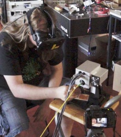
Our research group is conducting research in VR-OCT in close cooperation with physicians and engineers of the University of Lübeck. With a 1.6 MHz-FDML-Laser, 2 NVidia GeForce GTX1080-Ti and a specially developed imaging software up to 28 volumes per second can be achieved. This allows a smooth visualization of the rendered OCT volumes in a virtual environment, e.g. with the help of a HTC Vive.
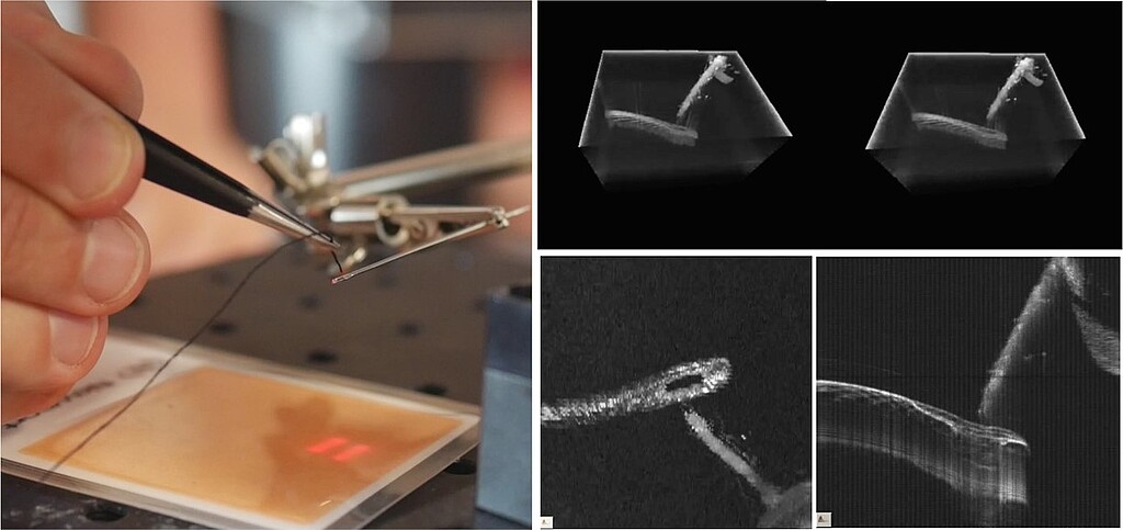
Video 1: Swine eye surgery using OCT in a virtual environment in real time (in cooperation with AG-Miura)
")


