Phase-sensitive optical coherence tomography (PhS-OCT)
OCT contrast technique based on phase information
The phase-sensitive optical coherence tomography (PhS-OCT) method is based on the same principles as optical coherence tomography (OCT). However, in PhS-OCT, the phase of the reflected light is extracted and analyzed in addition to the intensity of the light. When light interacts with biological tissue, changes in the optical path length and therefore the phase can occur due to variations in the structure and composition of the tissue. Consequently, the phase data can provide additional information about tissue changes, such as elasticity. An advantage here is that, due to the high sensitivity, even smallest changes in the sub-micrometer range can be detected and examined. In this way, tissue movements and differences that are barely or not at all noticeable in the intensity image can be visualized and evaluated. In addition, our high imaging speed can contribute significantly to the suppression of unwanted object or patient motion and is therefore of great interest.
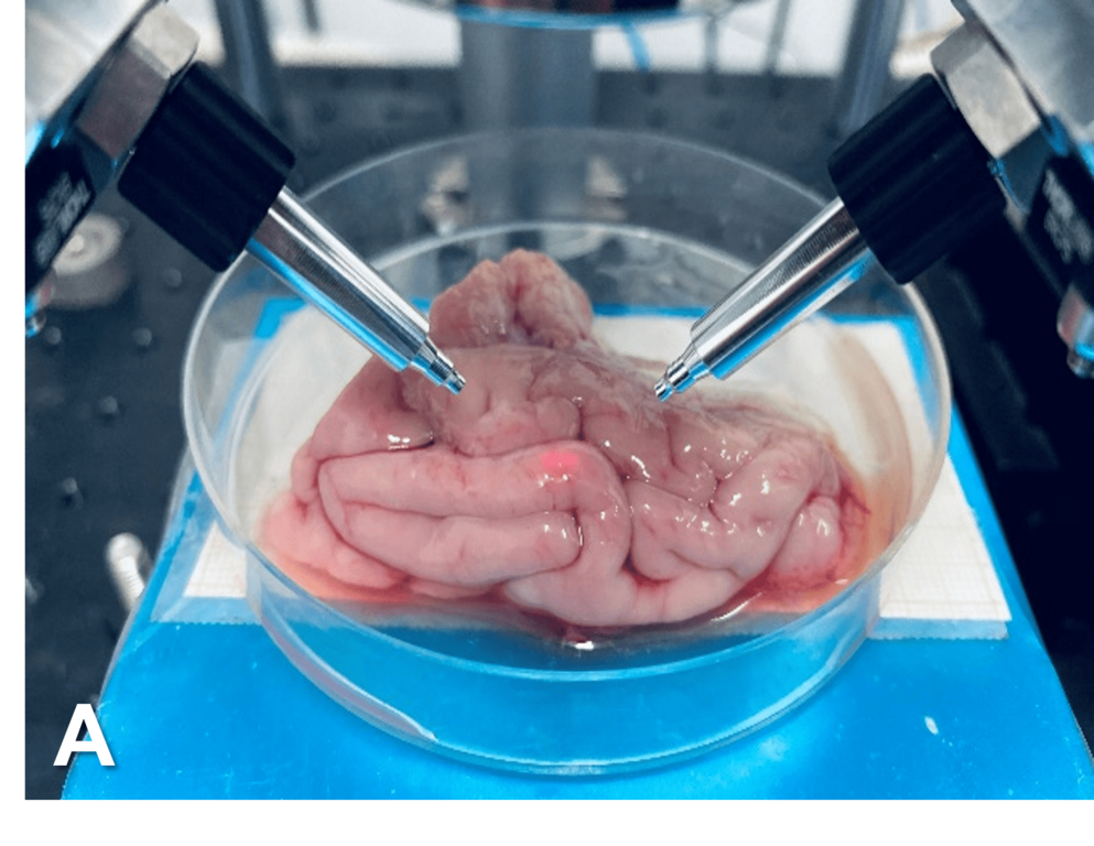
PhS-OCT has a wide range of applications in biomedical research and clinical practice, making it a valuable tool for studying the structure and function of biological tissue. The research group's current focus is on dermatology and neuro-oncology.