OCT is a noninvasive Imaging modality which is typically used for high resolution (~10µm), three dimensional imaging of scattering tissue. By using home built FDML laser technology we achieve imaging speeds of several million depth scans per second, which is one to two orders of magnitude higher than current commercially available systems (MHz-OCT).
These high imaging speeds already proved to be very useful in clinical applications, by reducing acquisition times and therefore reducing motion artifacts. But the high speed also gives access to the phase of the detected light and will thus allow the use of new numerical approaches for image quality enhancement and functional imaging with Swept-Source-OCT.
Our working group is conducting research in the field of OCT to develop new technologies and to identify possible fields of application.
The focus areas are:
- MHz-OCT - Ultra-fast OCT imaging with several million depth scans per second
- LARA-OCT - Large area robotically assisted OCT
- VR-OCT - Real-time computation and rendering of entire OCT volumes in a virtual environment
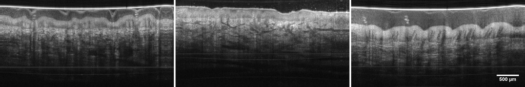
- Eye OCT - application of MHz-OCT to the eye for visualization of the retina or the anterior segment of the eye
- Phase sensitive OCT - enhancement of the information content of an OCT image by adding phase contrast
- Multispectral OCT - combination of RGB and OCT images for improved visualization of morphological structures
related Publications
2019
Zero roll-off retinal MHz-OCT using an FDML-laser, in Optical Coherence Imaging Techniques and Imaging in Scattering Media III , SPIE, Jul.2019. pp. 110780S.
| DOI: | 10.1117/12.2527034 |
| File: | 12.2527034.short |
| Bibtex: | @inproceedings{10.1117/12.2527034,
author = {Julian Klee and Jan Philip Kolb and Christin Grill and Wolfgang Draxinger and Tom Pfeiffer and Robert Huber},
title = {{Zero roll-off retinal MHz-OCT using an FDML-laser}},
volume = {11078},
booktitle = {Optical Coherence Imaging Techniques and Imaging in Scattering Media III},
editor = {Maciej Wojtkowski and Stephen A. Boppart and Wang-Yuhl Oh},
organization = {International Society for Optics and Photonics},
publisher = {SPIE},
pages = {110780S},
abstract = {Optical coherence tomography (OCT) applications like ultra-widefield and full eye-length imaging are of high interest for various diagnostic purposes. In swept-source OCT these techniques require a swept light source, which is coherent over the whole imaging depth. We present a zero roll-off 1060 nm Fourier Domain Mode Locked-Laser (FDML-Laser) for retinal OCT imaging at 1.7 MHz A-scan rate and first long-range imaging results with it. Several steps such as improved dispersion compensation and frequency regulation were performed and will be discussed. Besides virtually no loss in OCT signal over the maximum depth range of 4.6 mm and very good dynamic range was observed. Roll-off measurements show no decrease of the point-spread function (PSF), while maintaining a high dynamic range.},
keywords = {optical coherence tomography, OCT, tunable laser, Fourier Domain Mode Locking, FDML, MHz OCT},
year = {2019},
doi = {10.1117/12.2527034},
URL = {https://doi.org/10.1117/12.2527034}
} |
Live video rate volumetric OCT imaging of the retina with multi-MHz A-scan rates, PLOS ONE , vol. 14, no. 7, pp. e0213144, Mar. 2019.
| DOI: | 10.1371/journal.pone.0213144 |
| Bibtex: | @article{Kolb2019,
author = {Kolb, J P;Draxinger, W;Klee, J;Pfeiffer, T;Eibl, M;Klein, T;Wieser, W and Huber, R},
title = {Live video rate volumetric OCT imaging of the retina with multi-MHz A-scan rates},
journal = {J pone},
keywords = {AG-Huber_OCT},
url = {https://doi.org/10.1371/journal.pone.0213144},
pages = {e0213144},
ISSN = {1932-6203},
year = {2019},
type = {Journal Article}
}
|
A real-time video-rate 4D MHz-OCT microscope with high definition and low latency virtual reality display, in Optical Coherence Imaging Techniques and Imaging in Scattering Media III , Optica Publishing Group, 2019. pp. 11078_1.
| DOI: | 10.1117/12.2527177 |
Measurement of Inter-Sweep Phase Stability of an FDML Laser with a 10 kHz Tunable Ring Laser, in 2019 Conference on Lasers and Electro-Optics Europe and European Quantum Electronics Conference , Optical Society of America, 2019. pp. 1-1.
| DOI: | 10.1109/CLEOE-EQEC.2019.8872860 |
| Bibtex: | @inproceedings{Kastner:19,
author = {Kastner, D; Bl\"{o}mker, T; Pfeiffer, T; Grill, C; Schmidt, M; Jirauschek, C and Huber, R},
booktitle = {2019 Conference on Lasers and Electro-Optics Europe and European Quantum Electronics Conference},
journal = {2019 Conference on Lasers and Electro-Optics Europe and European Quantum Electronics Conference},
keywords = {Fourier domain mode locking; Image quality; Optical coherence tomography; Phase noise; Ring lasers; Tunable lasers},
pages = {cj_7_5},
publisher = {Optical Society of America},
title = {Measurement of Inter-Sweep Phase Stability of an FDML Laser with a 10 kHz Tunable Ring Laser},
year = {2019},
keywords = {AG-Huber_FDML, AG-Huber_OCT},
doi = { 10.1109/CLEOE-EQEC.2019.8872860},
abstract = {Fourier Domain Mode Locking (FDML) lasers are light sources that generate a sequence of narrowband optical frequency sweeps at the fundamental or harmonic of the cavity repetition rate \[1\]. This frequency swept output can also be considered as a sequence of strongly chirped, long pulses. FDML lasers are mainly used in swept source optical coherence tomography (SS-OCT), a medical imaging technique. The coherence length of the source, i.e. the intra-sweep phase stability of an FDML sweep, is decisive for the image quality and performance of OCT imaging \[2\].},
} |
2018
Combined in-depth, 3D, en face imaging of the optic disc, optic disc pits and optic disc pit maculopathy using swept-source megahertz OCT at 1050 nm, Graefes Arch Clin Exp Ophthalmol , vol. 256, no. 2, pp. 289-298, Dec. 2018.
| DOI: | 10.1007/s00417-017-3857-9 |
| Bibtex: | @article{Maertz2018,
author = {Maertz, J; Kolb, J P; Klein, T; Mohler, K J; Eibl, M; Wieser, W; Huber, R; Priglinger, S and Wolf, A},
title = {Combined in-depth, 3D, en face imaging of the optic disc, optic disc pits and optic disc pit maculopathy using swept-source megahertz OCT at 1050 nm},
journal = {Graefe's Archive for Clinical and Experimental Ophthalmology},
number = {2},
pages = {289-298},
DOI = {10.1007/s00417-017-3857-9},
url = {https://www.scopus.com/inward/record.uri?eid=2-s2.0-85032262413&doi=10.1007%2fs00417-017-3857-9&partnerID=40&md5=a46c315f12cf5e633ea0f7e644116eb3},
year = {2018},
Keywords= {En face imaging, Optical coherence tomography, Swept-source OCT, Megahertz OCT, 3D rendering, Optic disc, Optic disc pit, Optic disc pit maculopathy, AG-Huber_OCT},
type = {Journal Article}
} |
High-speed fiber scanning endoscope for volumetric multi-megahertz optical coherence tomography, Opt. Lett. , vol. 43, no. 18, pp. 4386-4389, Sep. 2018. Optica Publishing Group.
| DOI: | 10.1364/OL.43.004386 |
| Bibtex: | @article{Schulz-Hildebrandt:18,
author = {Hinnerk Schulz-Hildebrandt and Tom Pfeiffer and Tim Eixmann and Sabrina Lohmann and Martin Ahrens and Joshua Rehra and Wolfgang Draxinger and Peter K\"{o}nig and Robert Huber and Gereon H\"{u}ttmann},
journal = {Opt. Lett.},
keywords = {Fiber optics imaging; Endoscopic imaging; Medical and biological imaging; Optical coherence tomography; Fourier domain mode locking; Image quality; Optical coherence tomography; Single mode fibers; Step index fibers; Three dimensional imaging},
number = {18},
pages = {4386--4389},
publisher = {Optica Publishing Group},
title = {High-speed fiber scanning endoscope for volumetric multi-megahertz optical coherence tomography},
volume = {43},
month = {Sep},
year = {2018},
url = {https://opg.optica.org/ol/abstract.cfm?URI=ol-43-18-4386},
doi = {10.1364/OL.43.004386},
abstract = {We present a forward-viewing fiber scanning endoscope (FSE) for high-speed volumetric optical coherence tomography (OCT). The reduction in size of the probe was achieved by substituting the focusing optics by an all-fiber-based imaging system which consists of a combination of scanning single-mode fibers, a glass spacer, made from a step-index multi-mode fiber, and a gradient-index fiber. A lateral resolution of 11 $\mu$m was achieved at a working distance of 1.2 mm. The newly designed piezo-based FSE has an outer diameter of 1.6 mm and a rigid length of 13.5 mm. By moving the whole imaging optic in spirals for scanning the sample, the beam quality remains constant over the entire field of view with a diameter of 0.8 mm. The scanning frequency was adjusted to 1.22 kHz for use with a 3.28 MHz Fourier domain mode locked OCT system. Densely sampled volumes have been imaged at a rate of 6 volumes per second.},
} |
Ultra low noise Fourier domain mode locked laser for high quality megahertz optical coherence tomography, Biomed. Opt. Express , vol. 9, no. 9, pp. 4130-4148, Sep. 2018. Optica Publishing Group.
| DOI: | 10.1364/BOE.9.004130 |
| Bibtex: | @article{Pfeiffer:18,
author = {Tom Pfeiffer and Markus Petermann and Wolfgang Draxinger and Christian Jirauschek and Robert Huber},
journal = {Biomed. Opt. Express},
keywords = {Fiber optics imaging; Lasers, fiber; Optical coherence tomography; Laser stabilization ; Lasers, frequency modulated ; Analog to digital converters; Dark solitons; Image quality; Laser modes; Mode locking; Optical coherence tomography},
number = {9},
pages = {4130--4148},
publisher = {Optica Publishing Group},
title = {Ultra low noise Fourier domain mode locked laser for high quality megahertz optical coherence tomography},
volume = {9},
month = {Sep},
year = {2018},
url = {https://opg.optica.org/boe/abstract.cfm?URI=boe-9-9-4130},
doi = {10.1364/BOE.9.004130},
abstract = {We investigate the origin of high frequency noise in Fourier domain mode locked (FDML) lasers and present an extremely well dispersion compensated setup which virtually eliminates intensity noise and dramatically improves coherence properties. We show optical coherence tomography (OCT) imaging at 3.2 MHz A-scan rate and demonstrate the positive impact of the described improvements on the image quality. Especially in highly scattering samples, at specular reflections and for strong signals at large depth, the noise in optical coherence tomography images is significantly reduced. We also describe a simple model that suggests a passive physical stabilizing mechanism that leads to an automatic compensation of remaining cavity dispersion in FDML lasers.},
} |
High-resolution retinal swept source optical coherence tomography with an ultra-wideband Fourier-domain mode-locked laser at MHz A-scan rates, Biomed. Opt. Express , vol. 9, no. 1, pp. 120-130, Jan. 2018. Optica Publishing Group.
| DOI: | 10.1364/BOE.9.000120 |
| Bibtex: | @article{Kolb:18,
author = {Jan Philip Kolb and Tom Pfeiffer and Matthias Eibl and Hubertus Hakert and Robert Huber},
journal = {Biomed. Opt. Express},
keywords = {Medical optics instrumentation; Lasers, fiber; Medical and biological imaging; Ophthalmic optics and devices ; Optical coherence tomography; Adaptive optics; Image quality; In vivo imaging; Mode locking; Ophthalmic imaging; Three dimensional imaging},
number = {1},
pages = {120--130},
publisher = {Optica Publishing Group},
title = {High-resolution retinal swept source optical coherence tomography with an ultra-wideband Fourier-domain mode-locked laser at MHz A-scan rates},
volume = {9},
month = {Jan},
year = {2018},
url = {https://opg.optica.org/boe/abstract.cfm?URI=boe-9-1-120},
doi = {10.1364/BOE.9.000120},
abstract = {We present a new 1060 nm Fourier domain mode locked laser (FDML laser) with a record 143 nm sweep bandwidth at 2\&\#x2219;\&\#x202F;417 kHz\&\#x202F; $=$ \&\#x202F;834 kHz and 120 nm at 1.67 MHz, respectively. We show that not only the bandwidth alone, but also the shape of the spectrum is critical for the resulting axial resolution, because of the specific wavelength-dependent absorption of the vitreous. The theoretical limit of our setup lies at 5.9 \&\#x00B5;m axial resolution. In vivo MHz-OCT imaging of human retina is performed and the image quality is compared to the previous results acquired with 70 nm sweep range, as well as to existing spectral domain OCT data with 2.1 \&\#x00B5;m axial resolution from literature. We identify benefits of the higher resolution, for example the improved visualization of small blood vessels in the retina besides several others.},
} |
2017
Thermo-elastic optical coherence tomography, Optica Publishing Group, Sep.2017. pp. 3466-3469.
| DOI: | 10.1364/OL.42.003466 |
| Bibtex: | @article{Wang:17,
author = {Tianshi Wang and Tom Pfeiffer and Min Wu and Wolfgang Wieser and Gaetano Amenta and Wolfgang Draxinger and Antonius F. W. van der Steen and Robert Huber and Gijs van Soest},
journal = {Opt. Lett.},
keywords = {Imaging systems; Medical and biological imaging; Optical coherence tomography; Lasers, pulsed ; Fourier domain mode locking; Functional imaging; Laser beams; Nanosecond pulses; Optical coherence tomography; Phantom studies},
number = {17},
pages = {3466--3469},
publisher = {Optica Publishing Group},
title = {Thermo-elastic optical coherence tomography},
volume = {42},
month = {Sep},
year = {2017},
url = {https://opg.optica.org/ol/abstract.cfm?URI=ol-42-17-3466},
doi = {10.1364/OL.42.003466},
abstract = {The absorption of nanosecond laser pulses induces rapid thermo-elastic deformation in tissue. A sub-micrometer scale displacement occurs within a few microseconds after the pulse arrival. In this Letter, we investigate the laser-induced thermo-elastic deformation using a 1.5 MHz phase-sensitive optical coherence tomography (OCT) system. A displacement image can be reconstructed, which enables a new modality of phase-sensitive OCT, called thermo-elastic OCT. An analysis of the results shows that the optical absorption is a dominating factor for the displacement. Thermo-elastic OCT is capable of visualizing inclusions that do not appear on the structural OCT image, providing additional tissue type information.},
} |
1060nm FDML laser with centimeter coherence length and 1.67 MHz sweep rate for full eye length and retinal ultra-widefield OCT, in Optical Coherence Imaging Techniques and Imaging in Scattering Media II , Maciej Wojtkowski and Stephen A. Boppart and Wang-Yuhl Oh, Eds. SPIE, Aug.2017. pp. 104160J.
| DOI: | 10.1117/12.2286854 |
| Bibtex: | @inproceedings{10.1117/12.2286854,
author = {Jan Philip Kolb and Julian Klee and Tom Pfeiffer and Robert Huber},
title = {{1060nm FDML laser with centimeter coherence length and 1.67 MHz sweep rate for full eye length and retinal ultra-widefield OCT}},
volume = {10416},
booktitle = {Optical Coherence Imaging Techniques and Imaging in Scattering Media II},
editor = {Maciej Wojtkowski and Stephen A. Boppart and Wang-Yuhl Oh},
organization = {International Society for Optics and Photonics},
publisher = {SPIE},
pages = {104160J},
abstract = {We present a new design of a 1060nm Fourier Domain Mode Locked-Laser (FDML-Laser) that combines 1.67 MHz A-scan rate with a centimeter scale coherence length. The extended coherence length is achieved by synchronizing the cavity roundtrip time over the 75 nm sweep with a relative accuracy of 10<sup>-7</sup>. We will show that this requires careful combination of multiple fiber types in the cavity with a gradient heated chirped Fiber Bragg grating.},
keywords = {optical coherence tomograhy, OCT, tunable laser, Fourier domain mode locking, FDML, MHz OCT},
year = {2017},
doi = {10.1117/12.2286854},
URL = {https://doi.org/10.1117/12.2286854}
}
|
Long-range live 3D-OCT at different spectral zoom levels, in Optical Coherence Imaging Techniques and Imaging in Scattering Media II , Maciej Wojtkowski and Stephen A. Boppart and Wang-Yuhl Oh, Eds. SPIE, Aug.2017. pp. 104160L.
| DOI: | 10.1117/12.2287484 |
| Bibtex: | @inproceedings{10.1117/12.2287484,
author = {Tom Pfeiffer and Wolfgang Draxinger and Christin Grill and Robert Huber},
title = {{Long-range live 3D-OCT at different spectral zoom levels}},
volume = {10416},
booktitle = {Optical Coherence Imaging Techniques and Imaging in Scattering Media II},
editor = {Maciej Wojtkowski and Stephen A. Boppart and Wang-Yuhl Oh},
organization = {International Society for Optics and Photonics},
publisher = {SPIE},
pages = {104160L},
abstract = {We demonstrate that the 3.2 MHz a-scan rate and the improved coherence of our new low noise FDML laser enables live 3D-OCT with different spectral zooms and up to 10 cm of imaging range.},
keywords = {Optical coherence tomography, Fourier Domain Mode Locking, FDML, OCT},
year = {2017},
doi = {10.1117/12.2287484},
URL = {https://doi.org/10.1117/12.2287484}
}
|
INTRAPAPILLARY PROLIFERATION IN OPTIC DISK PITS: Clinical Findings and Time-Related Changes, Retina , vol. 37, no. 5, pp. 906-914, May 2017.
| DOI: | 10.1097/iae.0000000000001260 |
| Bibtex: | @article{Maertz2017,
author = {Maertz, J. and Mohler, K. J. and Kolb, J. P. and Klein, T. and Neubauer, A. and Kampik, A. and Priglinger, S. and Wieser, W. and Huber, R. and Wolf, A.},
title = {INTRAPAPILLARY PROLIFERATION IN OPTIC DISK PITS: Clinical Findings and Time-Related Changes},
journal = {Retina},
volume = {37},
number = {5},
pages = {906-914},
DOI = {10.1097/iae.0000000000001260},
year = {2017},
keywords = {AG-Huber_OCT},
type = {Journal Article}
}
|
Feature tracking for automated volume of interest stabilization on 4D-OCT images, in Medical Imaging 2017: Image-Guided Procedures, Robotic Interventions, and Modeling , Robert J. Webster III and Baowei Fei, Eds. SPIE, Mar.2017. pp. 101350W.
| DOI: | 10.1117/12.2255090 |
| Bibtex: | @inproceedings{10.1117/12.2255090,
author = {Max-Heinrich Laves and Andreas Schoob and L{\"u}der A. Kahrs and Tom Pfeiffer and Robert Huber and Tobias Ortmaier},
title = {{Feature tracking for automated volume of interest stabilization on 4D-OCT images}},
volume = {10135},
booktitle = {Medical Imaging 2017: Image-Guided Procedures, Robotic Interventions, and Modeling},
editor = {Robert J. Webster III and Baowei Fei},
organization = {International Society for Optics and Photonics},
publisher = {SPIE},
pages = {101350W},
abstract = {A common representation of volumetric medical image data is the triplanar view (TV), in which the surgeon manually selects slices showing the anatomical structure of interest. In addition to common medical imaging such as MRI or computed tomography, recent advances in the field of optical coherence tomography (OCT) have enabled live processing and volumetric rendering of four-dimensional images of the human body. Due to the region of interest undergoing motion, it is challenging for the surgeon to simultaneously keep track of an object by continuously adjusting the TV to desired slices. To select these slices in subsequent frames automatically, it is necessary to track movements of the volume of interest (VOI). This has not been addressed with respect to 4DOCT images yet. Therefore, this paper evaluates motion tracking by applying state-of-the-art tracking schemes on maximum intensity projections (MIP) of 4D-OCT images. Estimated VOI location is used to conveniently show corresponding slices and to improve the MIPs by calculating thin-slab MIPs. Tracking performances are evaluated on an in-vivo sequence of human skin, captured at 26 volumes per second. Among investigated tracking schemes, our recently presented tracking scheme for soft tissue motion provides highest accuracy with an error of under 2.2 voxels for the first 80 volumes. Object tracking on 4D-OCT images enables its use for sub-epithelial tracking of microvessels for image-guidance.},
keywords = {4D imaging, maximum intensity projection, optical coherence tomography, feature tracking},
year = {2017},
doi = {10.1117/12.2255090},
URL = {https://doi.org/10.1117/12.2255090}
}
|
Short pulse laser induced thermo-elastic deformation imaging, in Optical Interactions with Tissue and Cells XXVIII , E. Duco Jansen and Hope Thomas Beier, Eds. SPIE, Feb.2017. pp. 100620C.
| DOI: | 10.1117/12.2251502 |
| Bibtex: | @inproceedings{10.1117/12.2251502,
author = {Tianshi Wang and Tom Pfeiffer and Min Wu and Wolfgang Wieser and Wolfgang Draxinger and Antonius F. W. van der Steen and Robert Huber and Gijs van Soest},
title = {{Short pulse laser induced thermo-elastic deformation imaging}},
volume = {10062},
booktitle = {Optical Interactions with Tissue and Cells XXVIII},
editor = {E. Duco Jansen and Hope Thomas Beier},
organization = {International Society for Optics and Photonics},
publisher = {SPIE},
pages = {100620C},
abstract = {Absorption of nanosecond laser pulses induces rapid thermo-elastic deformation in tissue, i.e. a sub-micrometer scale displacement happens within a couple of microseconds. In this study, we initially investigate the depth-resolved deformation using a 1.5 MHz phase-sensitive optical coherence tomography (OCT) system. Functional images can be reconstructed based on the detected deformation, which enables a new imaging modality called thermo-elastic deformation imaging (TDI). Our results show that the associated displacement is related to the optical absorption of the short laser pulses. The TDI images can provide tissue type information in addition to the conventional OCT images.},
keywords = {thermal-elastic deformation, optical coherence tomography},
year = {2017},
doi = {10.1117/12.2251502},
URL = {https://doi.org/10.1117/12.2251502}
}
|
High-speed OCT light sources and systems [Invited], Biomed. Opt. Express , vol. 8, no. 2, pp. 828-859, Feb. 2017. Optica Publishing Group.
| DOI: | 10.1364/BOE.8.000828 |
| Bibtex: | @article{Klein:17,
author = {Thomas Klein and Robert Huber},
journal = {Biomed. Opt. Express},
keywords = {Imaging systems; Optical coherence tomography; Lasers and laser optics; Lasers, tunable; Optical coherence tomography; Full field optical coherence tomography; High speed imaging; Image quality; Imaging systems; Light wavelength; X ray imaging},
number = {2},
pages = {828--859},
publisher = {Optica Publishing Group},
title = {High-speed OCT light sources and systems \[Invited\]},
volume = {8},
month = {Feb},
year = {2017},
url = {https://opg.optica.org/boe/abstract.cfm?URI=boe-8-2-828},
doi = {10.1364/BOE.8.000828},
abstract = {Imaging speed is one of the most important parameters that define the performance of optical coherence tomography (OCT) systems. During the last two decades, OCT speed has increased by over three orders of magnitude. New developments in wavelength-swept lasers have repeatedly been crucial for this development. In this review, we discuss the historical evolution and current state of the art of high-speed OCT systems, with focus on wavelength swept light sources and swept source OCT systems.},
} |
Analysis of FDML lasers with meter range coherence, in Optical Coherence Tomography and Coherence Domain Optical Methods in Biomedicine XXI , James G. Fujimoto and Joseph A. Izatt and Valery V. Tuchin, Eds. SPIE, 2017. pp. 100531T.
| DOI: | 10.1117/12.2254792 |
| Bibtex: | @inproceedings{10.1117/12.2254792,
author = {Tom Pfeiffer and Wolfgang Draxinger and Wolfgang Wieser and Thomas Klein and Markus Petermann and Robert Huber},
title = {{Analysis of FDML lasers with meter range coherence}},
volume = {10053},
booktitle = {Optical Coherence Tomography and Coherence Domain Optical Methods in Biomedicine XXI},
editor = {James G. Fujimoto and Joseph A. Izatt and Valery V. Tuchin},
organization = {International Society for Optics and Photonics},
publisher = {SPIE},
pages = {100531T},
abstract = {FDML lasers provide sweep rates in the MHz range at wide optical bandwidths, making them ideal sources for high
speed OCT. Recently, at lower speed, ultralong-range swept-source OCT has been demonstrated using a tunable
vertical cavity surface emitting laser (VCSEL) and also using a Vernier-tunable laser. These sources provide relatively
high sweep rates and meter range coherence lengths. In order to achieve similar coherence, we developed an extremely
well dispersion compensated Fourier Domain Mode Locked (FDML) laser, running at 3.2 MHz sweep rate and 120 nm
spectral bandwidth. We demonstrate that this laser offers meter range coherence and enables volumetric long range OCT
of moving objects.},
keywords = {Optical coherence tomography, OCT, tunable laser, Fourier domain mode locking, FDML, MHz OCT},
year = {2017},
doi = {10.1117/12.2254792},
URL = {https://doi.org/10.1117/12.2254792}
}
|
2016
Heartbeat OCT and Motion-Free 3D In Vivo Coronary Artery Microscopy, JACC: Cardiovascular Imaging , vol. 9, no. 5, pp. 622-623, 2016.
| DOI: | 10.1016/j.jcmg.2015.08.010 |
| Bibtex: | @article{WANG2016622,
title = {Heartbeat OCT and Motion-Free 3D In Vivo Coronary Artery Microscopy},
journal = {JACC: Cardiovascular Imaging},
volume = {9},
number = {5},
pages = {622-623},
year = {2016},
issn = {1936-878X},
doi = {https://doi.org/10.1016/j.jcmg.2015.08.010},
url = {https://www.sciencedirect.com/science/article/pii/S1936878X15006713},
author = {Tianshi Wang and Tom Pfeiffer and Evelyn Regar and Wolfgang Wieser and Heleen {van Beusekom} and Charles T. Lancee and Geert Springeling and Ilona Krabbendam-Peters and Antonius F.W. {van der Steen} and Robert Huber and Gijs {van Soest}}
} |
Megahertz FDML laser with up to 143nm sweep range for ultrahigh resolution OCT at 1050nm, in Optical Coherence Tomography and Coherence Domain Optical Methods in Biomedicine XX , Joseph A. Izatt and James G. Fujimoto and Valery V. Tuchin, Eds. SPIE, 2016. pp. 969703.
| DOI: | 10.1117/12.2214758 |
| Bibtex: | @inproceedings{10.1117/12.2214758,
author = {Jan Philip Kolb and Thomas Klein and Matthias Eibl and Tom Pfeiffer and Wolfgang Wieser and Robert Huber},
title = {{Megahertz FDML laser with up to 143nm sweep range for ultrahigh resolution OCT at 1050nm}},
volume = {9697},
booktitle = {Optical Coherence Tomography and Coherence Domain Optical Methods in Biomedicine XX},
editor = {Joseph A. Izatt and James G. Fujimoto and Valery V. Tuchin},
organization = {International Society for Optics and Photonics},
publisher = {SPIE},
pages = {969703},
abstract = {We present a new design of a Fourier Domain Mode Locked laser (FDML laser), which provides a new record in sweep
range at ~1μm center wavelength: At the fundamental sweep rate of 2x417 kHz we reach 143nm bandwidth and 120nm
with 4x buffering at 1.67MHz sweep rate. The latter configuration of our system is characterized: The FWHM of the
point spread function (PSF) of a mirror is 5.6μm (in tissue). Human in vivo retinal imaging is performed with the MHz
laser showing more details in vascular structures. Here we could measure an axial resolution of 6.0μm by determining
the FWHM of specular reflex in the image. Additionally, challenges related to such a high sweep bandwidth such as
water absorption are investigated.},
keywords = {Optical coherence tomography, OCT, tunable laser, Fourier domain mode locking, FDML, MHz OCT},
year = {2016},
doi = {10.1117/12.2214758},
URL = {https://doi.org/10.1117/12.2214758}
}
|
2015
Heartbeat OCT: in vivo intravascular megahertz-optical coherence tomography, Biomed. Opt. Express , vol. 6, no. 12, pp. 5021-5032, Dec. 2015. Optica Publishing Group.
| DOI: | 10.1364/BOE.6.005021 |
| Bibtex: | @article{Wang:15,
author = {Tianshi Wang and Tom Pfeiffer and Evelyn Regar and Wolfgang Wieser and Heleen van Beusekom and Charles T. Lancee and Geert Springeling and Ilona Krabbendam and Antonius F.W. van der Steen and Robert Huber and Gijs van Soest},
journal = {Biomed. Opt. Express},
keywords = {Fiber optics imaging; Three-dimensional image acquisition; Medical optics instrumentation; Scanners; Endoscopic imaging; Medical and biological imaging; Optical coherence tomography; Image quality; Image registration; Imaging techniques; Laser modes; Mode locking; Optical coherence tomography},
number = {12},
pages = {5021--5032},
publisher = {Optica Publishing Group},
title = {Heartbeat OCT: in vivo intravascular megahertz-optical coherence tomography},
volume = {6},
month = {Dec},
year = {2015},
url = {https://opg.optica.org/boe/abstract.cfm?URI=boe-6-12-5021},
doi = {10.1364/BOE.6.005021},
abstract = {Cardiac motion artifacts, non-uniform rotational distortion and undersampling affect the image quality and the diagnostic impact of intravascular optical coherence tomography (IV-OCT). In this study we demonstrate how these limitations of IV-OCT can be addressed by using an imaging system that we called \&\#x201C;Heartbeat OCT\&\#x201D;, combining a fast Fourier Domain Mode Locked laser, fast pullback, and a micromotor actuated catheter, designed to examine a coronary vessel in less than one cardiac cycle. We acquired in vivo data sets of two coronary arteries in a porcine heart with both Heartbeat OCT, working at 2.88 MHz A-line rate, 4000 frames/s and 100 mm/s pullback speed, and with a commercial system. The in vivo results show that Heartbeat OCT provides faithfully rendered, motion-artifact free, fully sampled vessel wall architecture, unlike the conventional IV-OCT data. We present the Heartbeat OCT system in full technical detail and discuss the steps needed for clinical translation of the technology.},
} |
Combined 60° Wide-Field Choroidal Thickness Maps and High-Definition En Face Vasculature Visualization Using Swept-Source Megahertz OCT at 1050 nm60° High-Definition MHz-OCT Imaging of the Choroid, Investigative Ophthalmology & Visual Science , vol. 56, no. 11, pp. 6284--6293, Oct. 2015.
| DOI: | 10.1167/iovs.15-16670 |
| Bibtex: | @article{10.1167/iovs.15-16670,
author = {Mohler, Kathrin J. and Draxinger, Wolfgang and Klein, Thomas and Kolb, Jan Philip and Wieser, Wolfgang and Haritoglou, Christos and Kampik, Anselm and Fujimoto, James G. and Neubauer, Aljoscha S. and Huber, Robert and Wolf, Armin},
title = "{Combined 60° Wide-Field Choroidal Thickness Maps and High-Definition En Face Vasculature Visualization Using Swept-Source Megahertz OCT at 1050 nm}",
journal = {Investigative Ophthalmology & Visual Science},
volume = {56},
number = {11},
pages = {6284-6293},
year = {2015},
month = {10},
abstract = "{ To demonstrate ultrahigh-speed swept-source optical coherence tomography (SS-OCT) at 1.68 million A-scans/s for choroidal imaging in normal and diseased eyes over a ∼60° field of view. To investigate and correlate wide-field three-dimensional (3D) choroidal thickness (ChT) and vascular patterns using ChT maps and coregistered high-definition en face images extracted from a single densely sampled Megahertz-OCT (MHz-OCT) dataset. High-definition, ∼60° wide-field 3D datasets consisting of 2088 × 1024 A-scans were acquired using a 1.68 MHz prototype SS-OCT system at 1050 nm based on a Fourier-domain mode-locked laser. Nine subjects (nine eyes) with various chorioretinal diseases or without ocular pathology are presented. Coregistered ChT maps, choroidal summation maps, and depth-resolved en face images referenced to either the retinal pigment epithelium or the choroidal–scleral interface were generated using manual segmentation. Wide-field ChT maps showed a large inter- and intraindividual variance in peripheral and central ChT. In only four of the nine eyes, the location with the largest ChT was coincident with the fovea. The anatomy of the large lumen vessels of the outer choroid seems to play a major role in determining the global ChT pattern. Focal ChT changes with large thickness gradients were observed in some eyes. Different ChT and vascular patterns could be visualized over ∼60° in patients for the first time using OCT. Due to focal ChT changes, a high density of thickness measurements may be favorable. High-definition depth-resolved en face images are complementary to cross sections and thickness maps and enhance the interpretation of different ChT patterns. }",
issn = {1552-5783},
doi = {10.1167/iovs.15-16670},
url = {https://doi.org/10.1167/iovs.15-16670},
eprint = {https://arvojournals.org/arvo/content\_public/journal/iovs/934564/i1552-5783-56-11-6284.pdf},
} |
Wide-Field Megahertz OCT Imaging of Patients with Diabetic Retinopathy, Journal of Diabetes Research , vol. 2015, pp. 305084, Jul. 2015. Hindawi Publishing Corporation.
| DOI: | 10.1155/2015/305084 |
| Bibtex: | @article{Reznicek2015,
author = {Reznicek, Lukas and Kolb, Jan P. and Klein, Thomas and Mohler, Kathrin J. and Wieser, Wolfgang and Huber, Robert and Kernt, Marcus and Märtz, Josef and Neubauer, Aljoscha S.},
title = {Wide-Field Megahertz OCT Imaging of Patients with Diabetic Retinopathy},
journal = {Journal of Diabetes Research},
volume = {2015, Article ID 305084},
pages = {1-5},
DOI = {10.1155/2015/305084},
url = {http://dx.doi.org/10.1155/2015/305084},
year = {2015},
keywords = {AG-Huber_OCT},
type = {Journal Article}
}
|
High definition in vivo retinal volumetric video rate OCT at 0.6 Giga-voxels per second, in Optical Coherence Imaging Techniques and Imaging in Scattering Media , Brett E. Bouma and Maciej Wojtkowski, Eds. SPIE, Jul.2015. pp. 95410Z.
| DOI: | 10.1117/12.2183768 |
| Bibtex: | @inproceedings{10.1117/12.2183768,
author = {Jan Philip Kolb and Thomas Klein and Wolfgang Wieser and Wolfgang Draxinger and Robert Huber},
title = {{High definition in vivo retinal volumetric video rate OCT at 0.6 Giga-voxels per second}},
volume = {9541},
booktitle = {Optical Coherence Imaging Techniques and Imaging in Scattering Media},
editor = {Brett E. Bouma and Maciej Wojtkowski},
organization = {International Society for Optics and Photonics},
publisher = {SPIE},
pages = {95410Z},
abstract = {We present full volumetric high speed OCT imaging of the retina with multiple settings varying in volume size and volume rate. The volume size ranges from 255x255 A-scans to 160x40 A-scans with 450 samples per depth scan with volume rates varying between 20.8 V/s for the largest volumes to 195.2 V/s for the smallest. The system is based on a 1060nm Fourier domain mode locked (FDML) laser with 1.6MHz line rate. Scanning along the fast axis is performed with a 2.7 kHz or 4.3 kHz resonant scanner operated in bidirectional scanning mode, while a standard galvo scanner is used for the slow axis. The performance is analyzed with respect to various potential applications, like intraoperative OCT.},
keywords = {Optical coherence tomography, OCT, tunable laser, Fourier domain mode locking, FDML, MHz-OCT},
year = {2015},
doi = {10.1117/12.2183768},
URL = {https://doi.org/10.1117/12.2183768}
}
|
Fully automated 1.5 MHz FDML laser with more than 100mW output power at 1310 nm, in Optical Coherence Imaging Techniques and Imaging in Scattering Media , Brett E. Bouma and Maciej Wojtkowski, Eds. SPIE, Jul.2015. pp. 954116.
| DOI: | 10.1117/12.2183431 |
| Bibtex: | @inproceedings{10.1117/12.2183431,
author = {Wolfgang Wieser and Thomas Klein and Wolfgang Draxinger and Robert Huber},
title = {{Fully automated 1.5 MHz FDML laser with more than 100mW output power at 1310 nm}},
volume = {9541},
booktitle = {Optical Coherence Imaging Techniques and Imaging in Scattering Media},
editor = {Brett E. Bouma and Maciej Wojtkowski},
organization = {International Society for Optics and Photonics},
publisher = {SPIE},
pages = {954116},
abstract = {While FDML lasers with MHz sweep speeds have been presented five years ago, these devices have required manual control for startup and operation. Here, we present a fully self-starting and continuously regulated FDML laser with a sweep rate of 1.5 MHz. The laser operates over a sweep range of 115 nm centered at 1315 nm, and provides very high average output power of more than 100 mW. We characterize the laser performance, roll-off, coherence length and investigate the wavelength and phase stability of the laser output under changing environmental conditions. The high output power allows optical coherence tomography (OCT) imaging with an OCT sensitivity of 108 dB at 1.5 MHz.},
keywords = {OCT, optical coherence tomography, swept laser, wavelength-swept laser, fiber laser, MHz-OCT, Fourier-domain mode-locking, FDML},
year = {2015},
doi = {10.1117/12.2183431},
URL = {https://doi.org/10.1117/12.2183431}
} |
4-D Real-Time Optical Coherence Tomography, Opt. Photon. News , vol. 26, no. 6, pp. 32-39, Jun. 2015. Optica Publishing Group.
| DOI: | 10.1364/OPN.26.6.000032 |
| Bibtex: | @article{Huber:15,
author = {Robert Huber},
journal = {Opt. Photon. News},
keywords = {Image processing; Optical coherence tomography; Lasers, tunable; Medical optics and biotechnology; Optical coherence tomography; Image processing; Imaging techniques; Line scan cameras; Medical imaging; Optical coherence tomography; Three dimensional imaging},
number = {6},
pages = {32--39},
publisher = {Optica Publishing Group},
title = {4-D Real-Time Optical Coherence Tomography},
volume = {26},
month = {Jun},
year = {2015},
url = {https://www.optica-opn.org/abstract.cfm?URI=opn-26-6-32},
doi = {10.1364/OPN.26.6.000032},
abstract = {Advances in OCT techniques, combined with the processing power of moderncomputer hardware, are adding a new dimension---time---to a familiar 3-D imaging method.The result could be new applications in research and the biomedicalclinic.},
} |
")
Wolfgang Draxinger

AG Huber
Gebäude 81
,
Raum 72
wolfgang.draxinger(at)uni-luebeck.de
+49 451 3101 3229
Madita Göb

AG Huber
Gebäude 81
,
Raum 61
m.goeb(at)uni-luebeck.de
+49 451 3101 3262
Tjalfe Laedtke

AG Huber
Gebäude 81
,
Raum 61
tjalfe.laedtke(at)uni-luebeck.de
+49 451 3101 3265
Simon Lotz

AG Huber
Gebäude 81
,
Raum 72
si.lotz(at)uni-luebeck.de
+49 451 3101 3231


