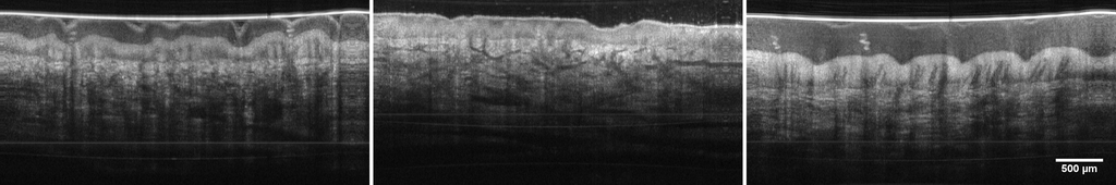
OCT is a noninvasive Imaging modality which is typically used for high resolution (~10µm), three dimensional imaging of scattering tissue. By using home built FDML laser technology we achieve imaging speeds of several million depth scans per second, which is one to two orders of magnitude higher than current commercially available systems (MHz-OCT).
These high imaging speeds already proved to be very useful in clinical applications, by reducing acquisition times and therefore reducing motion artifacts. But the high speed also gives access to the phase of the detected light and will thus allow the use of new numerical approaches for image quality enhancement and functional imaging with Swept-Source-OCT.
Our working group is conducting research in the field of OCT to develop new technologies and to identify possible fields of application.
The focus areas are:
- MHz-OCT - Ultra-fast OCT imaging with several million depth scans per second
- LARA-OCT - Large area robotically assisted OCT
- VR-OCT - Real-time computation and rendering of entire OCT volumes in a virtual environment
- Eye OCT - application of MHz-OCT to the eye for visualization of the retina or the anterior segment of the eye
- Phase sensitive OCT - enhancement of the information content of an OCT image by adding phase contrast
- Multispectral OCT - combination of RGB and OCT images for improved visualization of morphological structures
related Publications
2026
Processing pipeline for large optical coherence elastography datasets with quasi-static air-jet excitation: application to human brain tumor tissue, Biomed. Opt. Express , vol. 17, no. 3, pp. 1335—1358, Mar. 2026. Optica Publishing Group.
| DOI: | 10.1364/BOE.584263 |
| File: | abstract.cfm |
| Bibtex: | @article{Detrez:26,
author = {Nicolas Detrez and Sazgar Burhan and Jessica Kren and Jakob Matschke and Christian Hagel and Steffen Buschschl\"{u}ter and Dirk Theisen-Kunde and Matteo Mario Bonsanto and Robert Huber and Ralf Brinkmann},
journal = {Biomed. Opt. Express},
keywords = {Clinical applications; Deep learning; Elastography; Phase noise; Phase unwrapping; Tissue characterization},
number = {3},
pages = {1335--1358},
publisher = {Optica Publishing Group},
title = {Processing pipeline for large optical coherence elastography datasets with quasi-static air-jet excitation: application to human brain tumor tissue},
volume = {17},
month = {Mar},
year = {2026},
url = {https://opg.optica.org/boe/abstract.cfm?URI=boe-17-3-1335},
doi = {10.1364/BOE.584263},
abstract = {Optical coherence elastography (OCE) is a powerful imaging modality for assessing the mechanical properties of biological tissues. We employed an OCE system based on an Optores OMES 3.2 MHz OCT platform combined with an in-house developed air-jet excitation source to characterize healthy and tumorous (meningioma) human brain tissue. This paper presents a comprehensive software framework for processing large OCE datasets, enabling robust extraction of characteristic features from phase-derived displacement data and calculation of mechanical proxy parameters for detailed tissue characterization. Feature detection is achieved using a modified triangle threshold algorithm applied to the displacement curves from the OCE phase data. Extensive pre- and post-processing steps, including percentile-based filtering and adaptive histogram equalization, are applied to mitigate phase unwrapping errors and enhance visualization of the high dynamic range of OCE data. Exemplary measurements on human brain tumor samples demonstrate the framework's ability to differentiate between tissue types, highlighting its potential for future clinical and research applications.},
} |
A home-built flexible fiber laser to investigate optimal parameters for stimulating the tympanic membrane, in Optical Interactions with Tissue and Cells XXXVII , Joel N. Bixler and Alex J. Walsh and Norbert Linz, Eds. SPIE, 2026. pp. 1384904.
| DOI: | 10.1117/12.3080934 |
| Bibtex: | @inproceedings{10.1117/12.3080934,
author = {Henrik Volkens and Christin Grill and Florian Denk and Philipp Lamminger and Sebastian Freidank and Norbert Linz and Hendrik Husstedt and Robert Huber and Ralf Brinkmann},
title = {{A home-built flexible fiber laser to investigate optimal parameters for stimulating the tympanic membrane}},
volume = {13849},
booktitle = {Optical Interactions with Tissue and Cells XXXVII},
editor = {Joel N. Bixler and Alex J. Walsh and Norbert Linz},
organization = {International Society for Optics and Photonics},
publisher = {SPIE},
pages = {1384904},
abstract = {This work investigates optimizing optoacoustic stimulation of tympanic membrane models as a non-occlusive alternative to conventional acoustic drivers. We used a home-built, ytterbium-based master oscillator power amplifier (MOPA) operating at 1064 nm to stimulate an artificial tympanic membrane within a simplified middle ear model. The MOPA system can generate single laser pulses with 200 ps minimum pulse duration as well as concatenating multiple single pulses to MHz-bursts with burst durations up to 100 ns. Burst durations and burst energies were systematically varied between 30 and 100 ns and from 10 to 40 μJ. The laser-induced displacement of the membrane model was measured using phase-sensitive optical coherence tomography. Simultaneously the sound pressure level within a 0.4 ccm volume that mimics the middle ear cavity was measured. The results indicate that the membrane displacement and sound pressure increases both with higher burst energies at the same burst duration and longer burst durations at the same burst energy. Specifically, at a low burst repetition rate of 16 Hz, 100-ns pulse bursts yielded the most efficient stimulation. Furthermore, we demonstrated the system's capability for sound transmission up to 5 kHz by operating the MOPA at a repetition rate of 10 kHz. Using an acousto-optic modulator (AOM) for pulse amplitude modulation, we transmitted a speech signal onto the artificial membrane. The resulting acoustic signal was clearly audible and measurable within the middle ear model. These findings validate the feasibility of using tailored infrared laser pulses for middle ear stimulation. The ability to modulate complex audio signals via flexible, fiber-based laser architecture is a promising approach for developing next-generation hearing restoration technologies that avoid the occlusion effects and discomfort associated with traditional hearing aids.},
keywords = {Master oscillator fiber amplifier, Tympanic membrane, Temporal pulse shaping, Flexible fiber laser, Thermoelastic bending, Parameter optimization, Optical tissue stimulation},
year = {2026},
doi = {10.1117/12.3080934},
URL = {https://doi.org/10.1117/12.3080934}
}
|
Dynamic shockwave photography using a home-built MOFA laser system with flexible repetition rate up to 5 GHz, in Optical Interactions with Tissue and Cells XXXVII , Joel N. Bixler and Alex J. Walsh and Norbert Linz, Eds. SPIE, 2026. pp. PC1384903.
| DOI: | 10.1117/12.3080401 |
| Bibtex: | @inproceedings{10.1117/12.3080401,
author = {Henrik Volkens and Sebastian Freidank and Philipp Lamminger and Alfred Vogel and Robert Huber and Ralf Brinkmann and Norbert Linz},
title = {{Dynamic shockwave photography using a home-built MOFA laser system with flexible repetition rate up to 5 GHz}},
volume = {PC13849},
booktitle = {Optical Interactions with Tissue and Cells XXXVII},
editor = {Joel N. Bixler and Alex J. Walsh and Norbert Linz},
organization = {International Society for Optics and Photonics},
publisher = {SPIE},
pages = {PC1384903},
abstract = {Laser-induced ablation in liquids (LAL) is widely used for nanoparticle generation, yet its underlying mechanisms are not completely understood. We investigate interactions between shockwave, cavitation bubble and target material by multi exposure imaging with high temporal and spatial resolution. Our home-built Yb-based master oscillator fiber amplifier system delivers 170 ps pulses at 2 µJ and tunable burst rates up to 5 GHz, ideal for capturing transient events. Speckle-free imaging is achieved using a fiber-based rapid optical mode mixing approach combining spectral broadening with optical delay and spatial mode mixing of frequency-doubled 532 nm pulses.},
keywords = {Laser Ablation in Liquids (LAL), Shockwave Photography, High-Speed Imaging, Multi-Exposure Illumination, Master Oscillator Fiber Amplifier (MOFA), Speckle-Free Imaging, Cavitation Bubble, Nanoparticle Generation},
year = {2026},
doi = {10.1117/12.3080401},
URL = {https://doi.org/10.1117/12.3080401}
} |
Towards optoacoustic transient shaping using a flexible fiber laser system, in Photons Plus Ultrasound: Imaging and Sensing 2026 , Alexander A. Oraevsky and Lihong V. Wang, Eds. SPIE, 2026. pp. 138511F.
| DOI: | 10.1117/12.3080520 |
| Bibtex: | @inproceedings{10.1117/12.3080520,
author = {Henrik Volkens and Philipp Lamminger and Norbert Linz and Sebastian Freidank and Robert Huber and Ralf Brinkmann},
title = {{Towards optoacoustic transient shaping using a flexible fiber laser system}},
volume = {13851},
booktitle = {Photons Plus Ultrasound: Imaging and Sensing 2026},
editor = {Alexander A. Oraevsky and Lihong V. Wang},
organization = {International Society for Optics and Photonics},
publisher = {SPIE},
pages = {138511F},
abstract = {We aim to increase the efficiency of optoacoustic signal generation for precise, in vivo, real-time tissue temperature monitoring during thermal retinal interventions, by matching the timing of multiple laser excitation events to the acoustic response of the examined specimen. To achieve this goal, we utilized a home-built Ytterbium-based master oscillator power amplifier (MOPA) fiber laser system that provides unprecedented control over the temporal pulse structure, allowing for pulse-burst durations from picoseconds to nanoseconds and arbitrary repetition rates for investigating the influence of the excitation duration on the amplitude of the resulting optoacoustic transients. Methodologically, experiments were performed on ex vivo explants of porcine retinal pigment epithelium (RPE) consisting of the RPE, choroid, and sclera embedded in a cuvette filled with saline solution. Optoacoustic transients were detected using a piezoelectric ring transducer (fres = 1 MHz, Medical Laser Center Lübeck, Germany) integrated into a standard ophthalmic contact glass with a distance of 24 mm to the specimen. We systematically investigated the influence of pulse-burst durations between 10 and 100-ns with the total burst energy of 3 μJ matching a typical probe pulse energy. Each burst was produced with a repetition rate of 500 MHz. Results demonstrate that, at typical pulse energies of 3 μJ, shorter pulse-burst durations down to 30 ns significantly increase the amplitude of the generated acoustic transients compared to longer pulse-bursts. While higher burst energy consistently results in stronger signals, signal generation efficiency is highly dependent on the temporal burst width. With decreasing burst durations, the amplitude of the resulting transients decreases lower than that of the 30-ns burst. We hypothesize that shorter excitation bursts result in a signal consisting of higher-frequency components that are stronger attenuated in water. These findings highlight that tailoring the temporal excitation profile is essential for maximizing signal-to-noise ratio. The compact and scalable fiber-based MOPA architecture offers a versatile alternative to traditional bulk lasers, providing the necessary degrees of freedom for optimized optoacoustic tissue characterization and in future real-time monitoring.},
keywords = {Master Oscillator Fiber Amplifier (MOFA), Optoacoustics, Transient shaping, Temperature measurement, Tailored optoacoustic excitation, Flexible fiber laser, Retinal laser treatment, Multi-GHz repetition rate},
year = {2026},
doi = {10.1117/12.3080520},
URL = {https://doi.org/10.1117/12.3080520}
} |
Dual-resolution megahertz optical coherence tomography prototype rectoscope for enhanced visualization of colorectal microstructures, Journal of Biomedical Optics , vol. 31, no. 4, pp. 046002, 2026. SPIE.
| DOI: | 10.1117/1.JBO.31.4.046002 |
| Bibtex: | @article{10.1117/1.JBO.31.4.046002,
author = {Sazgar Burhan and Berenice Schulte and Madita G{\"o}b and Awanish Pratap Singh and Bayan Mustafa and Simon Lotz and Wolfgang Draxinger and Philipp Lamminger and Yasmeine Saker and Tim Eixmann and Martin Ahrens and Marvin Heimke and Tillmann Heinze and Thilo Wedel and Maik Rahlves and Mark Ellrichmann and Robert Huber},
title = {{Dual-resolution megahertz optical coherence tomography prototype rectoscope for enhanced visualization of colorectal microstructures}},
volume = {31},
journal = {Journal of Biomedical Optics},
number = {4},
publisher = {SPIE},
pages = {046002},
keywords = {MHz optical coherence tomography, rectoscope, dual mode design, extended-range imaging mode, high-detail imaging mode, ethanol-glycerol-lysoformin fixation, Optical coherence tomography, Imaging systems, Tissues, Rectum, Visualization, Biological imaging, GRIN lenses, Anatomy, Endoscopy, Image resolution},
year = {2026},
doi = {10.1117/1.JBO.31.4.046002},
URL = {https://doi.org/10.1117/1.JBO.31.4.046002}
} |
2025
Flow-Controlled Air-Jet for In Vivo Quasi Steady-State and Dynamic Elastography With MHz Optical Coherence Tomography, IEEE Transactions on Biomedical Engineering , vol. 72, no. 3, pp. 1008-1020, Mar. 2025.
| DOI: | 10.1109/TBME.2024.3484676 |
| Bibtex: | @ARTICLE{10726870,
author={Detrez, Nicolas and Burhan, Sazgar and Rewerts, Katarina and Kren, Jessica and Buschschlüter, Steffen and Theisen-Kunde, Dirk and Bonsanto, Matteo Mario and Huber, Robert and Brinkmann, Ralf},
journal={IEEE Transactions on Biomedical Engineering},
title={Flow-controlled air-jet for in vivo quasi steady-state and dynamic elastography with MHz optical coherence tomography},
year={2024},
volume={},
number={},
pages={1-12},
keywords={Force;Biomedical measurement;Pressure measurement;In vivo;Steady-state;Generators;Elastography;Valves;Force measurement;Optical coherence tomography;Air-Jet;Air-Puff;Optical Coherence Elastography;Stiffness;Tissue Mechanics;Young's Modulus},
doi={10.1109/TBME.2024.3484676}} |
Large-area dynamic contrast MHz optical coherence tomography for label-free imaging of porcine tissue, in Optical Coherence Tomography and Coherence Domain Optical Methods in Biomedicine XXIX , Rainer A. Leitgeb and Yoshiaki Yasuno, Eds. SPIE, Mar.2025. pp. 1330502.
| DOI: | 10.1117/12.3046216 |
| Weblink: | https://zenodo.org/records/19853630 |
| Bibtex: | @inproceedings{10.1117/12.3046216,
author = {Sazgar Burhan and Madita G{\"o}b and Mario Pieper and Tjalfe Laedtke and Thorge Grahl and Michael M{\"u}nter and Hinnerk Schulz-Hildebrandt and Gereon H{\"u}ttmann and Peter K{\"o}nig and Robert Huber},
title = {{Large-area dynamic contrast MHz optical coherence tomography for label-free imaging of porcine tissue}},
volume = {13305},
booktitle = {Optical Coherence Tomography and Coherence Domain Optical Methods in Biomedicine XXIX},
editor = {Rainer A. Leitgeb and Yoshiaki Yasuno},
organization = {International Society for Optics and Photonics},
publisher = {SPIE},
pages = {1330502},
abstract = {We demonstrate a 3.2 MHz-OCT system for inter-volumetric dynamic optical coherence tomography of ex vivo porcine kidney tissue. Employing a home-built Fourier Domain mode locking (FDML) laser with a 1310 nm wavelength, the system achieved a lateral resolution of 3.48 μm and a frame rate of 612 Hz. A motorized XYZ positioning stage enabled the precise acquisition of multiple volumes, which were seamlessly stitched together to generate a comprehensive dataset with a total area of 2.6 × 2.6 mm<sup>2</sup>. Validations against histological sections confirmed the system’s ability to visualize cellular tissue structures.},
keywords = {Optical Coherence Tomography, Megahertz OCT, Fourier Domain Mode Locking, Dynamic OCT, Functional OCT, Three-dimensional image acquisition, Tissue Dynamics, Kidney},
year = {2025},
doi = {10.1117/12.3046216},
URL = {https://doi.org/10.1117/12.3046216}
} |
Switchable lateral resolution real-time MHz-OCT rectoscopy for enhanced colorectal disease diagnosis, in Optical Coherence Tomography and Coherence Domain Optical Methods in Biomedicine XXIX , Rainer A. Leitgeb and Yoshiaki Yasuno, Eds. SPIE, Mar.2025. pp. 1330512.
| DOI: | 10.1117/12.3046222 |
| Bibtex: | @inproceedings{10.1117/12.3046222,
author = {Sazgar Burhan and Berenice Schulte and Madita G{\"o}b and Awanish Pratap Singh and Bayan Mustafa and Simon Lotz and Wolfgang Draxinger and Philipp Lamminger and Yasmeine Saker and Tim Eixmann and Martin Ahrens and Marvin Heimke and Tillmann Heinze and Thilo Wedel and Maik Rahlves and Mark Ellrichmann and Robert Huber},
title = {{Switchable lateral resolution real-time MHz-OCT rectoscopy for enhanced colorectal disease diagnosis}},
volume = {13305},
booktitle = {Optical Coherence Tomography and Coherence Domain Optical Methods in Biomedicine XXIX},
editor = {Rainer A. Leitgeb and Yoshiaki Yasuno},
organization = {International Society for Optics and Photonics},
publisher = {SPIE},
pages = {1330512},
abstract = {Endoscopic optical coherence tomography (OCT) offers in vivo live visualization of transmural structures with histological resolution, making it a valuable tool in medical imaging. In gastroenterology, OCT endoscopy is particularly advantageous for assessing rectal wall layers, providing superior axial and lateral resolution compared to conventional rectal endoscopic ultrasound. However, the large diameter and uneven colon surface present challenges for comprehensive imaging. Extending the OCT imaging range addresses this issue by enabling a thorough examination of the entire colon, facilitating the detection of surface polyps, tumors, and their infiltration depth. Once these regions of interest are identified, high-resolution imaging becomes essential for detailed evaluation. To meet these demands, this study integrates two different imaging modes, an extended-range mode, and a high-detail mode, within a rigid rectoscope. The extended-range mode enables visualization of deeper structures, while the high-detail mode enhances image quality for precise, contact-based assessments. The system allows seamless, real-time transitions between the modes using a 3.2MHz-OCT system and a fiber‑optic MEMS switch.},
keywords = {Optical Coherence Tomography, Megahertz OCT, Fourier Domain Mode Locking, Three-dimensional image acquisition, Rectal Imaging, Long-Range Imaging, Non-Invasive Diagnostic Imaging, Tumor Assessment},
year = {2025},
doi = {10.1117/12.3046222},
URL = {https://doi.org/10.1117/12.3046222}
} |
1.7MHz, 840nm swept-source ophthalmic OCT, in Ophthalmic Technologies XXXV , Daniel X. Hammer and Derek Nankivil and Yuankai K. Tao, Eds. SPIE, Mar.2025. pp. 1330004.
| DOI: | 10.1117/12.3045055 |
| Bibtex: | @inproceedings{10.1117/12.3045055,
author = {Marie Klufts and Wolfgang Draxinger and Simon Lotz and Robert Huber},
title = {{1.7MHz, 840nm swept-source ophthalmic OCT}},
volume = {13300},
booktitle = {Ophthalmic Technologies XXXV},
editor = {Daniel X. Hammer and Derek Nankivil and Yuankai K. Tao},
organization = {International Society for Optics and Photonics},
publisher = {SPIE},
pages = {1330004},
keywords = {swept source, SS-OCT, FDML , Retinal imaging, ophthalmic imaging, OCT, 850 nm, short wavelength},
year = {2025},
doi = {10.1117/12.3045055},
URL = {https://doi.org/10.1117/12.3045055}
} |
Towards ultrahigh resolution MHz retinal SS-OCT: 187nm section-wise tuning of a FDML laser at 1050nm, in Optical Coherence Tomography and Coherence Domain Optical Methods in Biomedicine XXIX , Rainer A. Leitgeb and Yoshiaki Yasuno, Eds. SPIE, Mar.2025. pp. 133050K.
| DOI: | 10.1117/12.3046386 |
| Bibtex: | @inproceedings{10.1117/12.3046386,
author = {M. A. Bashir and M. Klufts and S. Lotz and R. Huber},
title = {{Towards ultrahigh resolution MHz retinal SS-OCT: 187nm section-wise tuning of a FDML laser at 1050nm}},
volume = {13305},
booktitle = {Optical Coherence Tomography and Coherence Domain Optical Methods in Biomedicine XXIX},
editor = {Rainer A. Leitgeb and Yoshiaki Yasuno},
organization = {International Society for Optics and Photonics},
publisher = {SPIE},
pages = {133050K},
,
keywords = {wavelength-swept laser, FDML lasers, Optical coherence tomography, Fourier domain mode locked lasers, Broadband lasers, tunable lasers, swept lasers, swept source OCT},
year = {2025},
doi = {10.1117/12.3046386},
URL = {https://doi.org/10.1117/12.3046386}
} |
Enhancing brain tumor detection using optical coherence tomography and variational autoencoders, in Medical Imaging 2025: Clinical and Biomedical Imaging , Barjor S. Gimi and Andrzej Krol, Eds. SPIE, 2025. pp. 134101P.
| DOI: | 10.1117/12.3047226 |
| Bibtex: | @inproceedings{10.1117/12.3047226,
author = {Paul Strenge and Birgit Lange and Wolfgang Draxinger and Dirk Theisen-Kunde and Sonja Spahr-Hess and Matteo M. Bonsanto and Robert Huber and Ralf Brinkmann and Heinz Handels},
title = {{Enhancing brain tumor detection using optical coherence tomography and variational autoencoders}},
volume = {13410},
booktitle = {Medical Imaging 2025: Clinical and Biomedical Imaging},
editor = {Barjor S. Gimi and Andrzej Krol},
organization = {International Society for Optics and Photonics},
publisher = {SPIE},
pages = {134101P},
abstract = {Neurosurgical intervention is critical in brain tumor treatment, with long-term survival closely linked to the extent of tumor resection. The goal is to completely remove tumor tissue while preserving healthy tissue, a challenging task due to the diffuse nature of some brain tumors, such as glioblastoma, which infiltrate healthy tissue in ways that are difficult to distinguish histologically. Current intraoperative imaging techniques, including MRI and fluorescence microscopy, are limited in reliably identifying tumor tissue. Optical coherence tomography (OCT) offers a promising alternative, providing non-invasive, high-resolution cross-sectional images. This study investigates the use of a variational autoencoder (VAE) in combination with an evidential learning framework to enhance the classification of brain tissues in OCT images. The classification approach, applied to ex vivo OCT images captured at a wavelength of 1300 nm, achieved an average precision of 0.87 and a recall of 0.88 for the discrimination of healthy and tumorous brain tissue with consideration of prediction uncertainties. This method demonstrated improved discrimination between healthy white matter and tumor-infiltrated white matter compared to previous studies.},
keywords = {brain tumor, OCT, variational autoencoders, glioblastoma, classification, medical imaging, brain, evidential learning},
year = {2025},
doi = {10.1117/12.3047226},
URL = {https://doi.org/10.1117/12.3047226}
} |
Ultrashort Power-Dips in Fourier Domain Mode-Locked Lasers: Impact of Picosecond Carrier Recovery, in 2025 Conference on Lasers and Electro-Optics Europe & European Quantum Electronics Conference (CLEO/Europe-EQEC) , Jun.2025. pp. 1-1.
| DOI: | 10.1109/CLEO/Europe-EQEC65582.2025.11109071 |
| Bibtex: | @INPROCEEDINGS{11109071,
author={Aşırım, Özüm Emre and Huber, Robert and Jirauschek, Christian},
booktitle={2025 Conference on Lasers and Electro-Optics Europe & European Quantum Electronics Conference (CLEO/Europe-EQEC)},
title={Ultrashort Power-Dips in Fourier Domain Mode-Locked Lasers: Impact of Picosecond Carrier Recovery},
year={2025},
volume={},
number={},
pages={1-1},
abstract={Fourier domain mode-locked (FDML) lasers are widely used in applications requiring high-speed wavelength sweeps and reliable spectral stability, such as optical coherence tomography (OCT) [1]. In this study, we explore the steady-state behavior of FDML lasers when the carrier lifetime of the semiconductor optical amplifier (SOA) is reduced to one picosecond-a scenario that can enable reduced intensity noise, improved coherence, and higher sweep speed, achievable with advanced quantum-well or quantum-dot SOAs, opening possibilities for next-generation FDML lasers [2], [3]. In previous studies, SOA carrier lifetimes longer than 70 picoseconds yielded irregular dips (holes) with varying shape, amplitude, and duration in the output power pattern, hindering beam coherence except under ultra-stable conditions [1,3-5]. Our latest simulations, which align with experimental findings in the detection and profiling of such dips [3]–[5], reveal that a 1 ps carrier lifetime improves stability and coherence but leads to emergence of consistent ultrashort, sinc-like power dips (Fig. 1, middle) for high output powers, which are almost uniform in duration (Fig. 1, right). The density of these dips increases as the output power is raised toward the upper practical limit. Based on foundational FDML laser theory [5]–[6], Equations (1)-(3) explain the formation of these ultrafast dips. At high photon flux, rapid gain depletion causes a sharp drop in carrier density $(N)$, generating a dip. Given the ultrashort carrier lifetime $(\tau_{c})$, the carriers recover quickly after depletion, restoring gain for the next dip. The time-delayed feedback term in Equation (2) represents light from previous round trips interacting with the restored carriers, amplifying the dips.},
keywords={Semiconductor optical amplifiers;Laser mode locking;Power amplifiers;Coherence;Laser feedback;Laser stability;Stability analysis;Mathematical models;Charge carrier lifetime;Power generation},
doi={10.1109/CLEO/Europe-EQEC65582.2025.11109071},
ISSN={2833-1052},
month={June},} |
Megahertz dynamic optical coherence tomography of blisters in human skin, Biomed. Opt. Express , vol. 16, no. 10, pp. 4063—4078, Oct. 2025. Optica Publishing Group.
| DOI: | 10.1364/BOE.571621 |
| File: | abstract.cfm |
| Bibtex: | @article{Gob:25,
author = {Madita G\"{o}b and Linh Ha-Wissel and Caren Jacobi and Jennifer E. Hundt and Robert Huber},
journal = {Biomed. Opt. Express},
keywords = {Effective refractive index; High speed imaging; Image metrics; Imaging systems; Imaging techniques; In vivo imaging},
number = {10},
pages = {4063--4078},
publisher = {Optica Publishing Group},
title = {Megahertz dynamic optical coherence tomography of blisters in human skin},
volume = {16},
month = {Oct},
year = {2025},
url = {https://opg.optica.org/boe/abstract.cfm?URI=boe-16-10-4063},
doi = {10.1364/BOE.571621},
abstract = {Detecting epidermal blisters in human skin using optical coherence tomography (OCT) is clinically valuable, particularly for diagnosing autoimmune blistering diseases. Dynamic OCT (dOCT) extends conventional structural imaging by providing motion-based contrast sensitive to tissue dynamics. In this study, we analyze the diagnostic potential of dynamic contrast in high-speed (MHz-OCT) and microscopic (mOCT) OCT for blister imaging. We first evaluate whether these systems offer sufficient structural detail for blister detection, comparing them to a clinical reference. Dynamic contrast was then examined in an ex vivo human skin blister model using mOCT, and in vivo, MHz-OCT was subsequently applied to healthy and blistered skin. Our findings demonstrate improved layer delineation and blister localization. We further discuss system-specific image characteristics, artifacts, and their implications for future OCT-based diagnostic workflows.},
} |
Label-free volumetric imaging of porcine kidney tissue over extended areas using dynamic MHz-OCT, Scientific Reports , vol. 15, no. 1, pp. 32426, Sep. 2025.
| DOI: | 10.1038/s41598-025-15032-6 |
| File: | s41598-025-15032-6 |
| Bibtex: | @article{RN5536,
author = {Burhan, Sazgar;Göb, Madita;Pieper, Mario;Laedtke, Tjalfe;Grahl, Thorge;Münter, Michael;Schulz-Hildebrandt, Hinnerk;Hüttmann, Gereon;König, Peter and Huber, Robert},
title = {Label-free volumetric imaging of porcine kidney tissue over extended areas using dynamic MHz-OCT},
journal = {Scientific Reports},
volume = {15},
number = {1},
pages = {32426},
ISSN = {2045-2322},
DOI = {10.1038/s41598-025-15032-6},
url = {https://doi.org/10.1038/s41598-025-15032-6},
year = {2025},
type = {Journal Article}
} |
Lifting constraints on multi-kHz raster-line scanning frequency matching in multi-MHz Swept-Source OCT imaging systems, in European Conferences on Biomedical Optics 2025 , Optica Publishing Group, 2025. pp. W5D.5.
| DOI: | 10.1364/ECBO.2025.W5D.5 |
| File: | abstract.cfm |
| Bibtex: | @inproceedings{Draxinger:25,
author = {Wolfgang Draxinger and Simon Lotz and Allegra Behr and Madita G\"{o}b and Robert Huber},
booktitle = {European Conferences on Biomedical Optics 2025},
journal = {European Conferences on Biomedical Optics 2025},
keywords = {Absolute distance measurement; Field programmable gate arrays; Imaging systems; Light sources; Scanners; Swept sources},
pages = {W5D.5},
publisher = {Optica Publishing Group},
title = {Lifting constraints on multi-kHz raster-line scanning frequency matching in multi-MHz Swept-Source OCT imaging systems},
year = {2025},
url = {https://opg.optica.org/abstract.cfm?URI=ECBO-2025-W5D.5},
doi = {10.1364/ECBO.2025.W5D.5},
abstract = {The established synchronization scheme of SS-OCT calls for the raster-line frequency to be a remainder-less divider of the sweep frequency. Two methods are presented that increase flexibility in scanner operation.},
} |
Speckle Reduction Through Angular Compounding in Robotically Assisted MHz-OCT, in European Conferences on Biomedical Optics 2025 , Optica Publishing Group, 2025. pp. W1D.4.
| DOI: | 10.1364/ECBO.2025.W1D.4 |
| Weblink: | https://zenodo.org/records/19853892 |
| File: | abstract.cfm |
| Bibtex: | @inproceedings{Laedtke:25,
author = {Tjalfe Laedtke and Sazgar Burhan and Simon Lotz and Madita G\"{o}b and Robert Huber},
booktitle = {European Conferences on Biomedical Optics 2025},
journal = {European Conferences on Biomedical Optics 2025},
keywords = {Image registration; Imaging systems; Phase shift; Spatial resolution; Speckle noise; Speckle patterns},
pages = {W1D.4},
publisher = {Optica Publishing Group},
title = {Speckle Reduction Through Angular Compounding in Robotically Assisted MHz-OCT},
year = {2025},
url = {https://opg.optica.org/abstract.cfm?URI=ECBO-2025-W1D.4},
doi = {10.1364/ECBO.2025.W1D.4},
abstract = {We demonstrate speckle reduction in robotically assisted MHz-OCT by angular compounding. The robot is used to acquire images from different angles, which, after registration, are used for efficient speckle averaging without loss of spatial resolution.},
} |
Megahertz FDML laser with on-the-fly adjustable sweep rate between 835 kHz and 13.4 MHz, in European Conferences on Biomedical Optics 2025 , Optica Publishing Group, 2025. pp. W5D.4.
| DOI: | 10.1364/ECBO.2025.W5D.4 |
| File: | abstract.cfm |
| Bibtex: | @inproceedings{Lotz:25,
author = {Simon Lotz and Wolfgang Draxinger and Anneli Dick and Robert Huber},
booktitle = {European Conferences on Biomedical Optics 2025},
journal = {European Conferences on Biomedical Optics 2025},
keywords = {Fiber Bragg gratings; Imaging techniques; Laser sources; Optical buffers; Swept sources; Three dimensional imaging},
pages = {W5D.4},
publisher = {Optica Publishing Group},
title = {Megahertz FDML laser with on-the-fly adjustable sweep rate between 835 kHz and 13.4 MHz},
year = {2025},
url = {https://opg.optica.org/abstract.cfm?URI=ECBO-2025-W5D.4},
doi = {10.1364/ECBO.2025.W5D.4},
abstract = {We present a Megahertz FDML laser which can be automatically, and on-the-fly switched to speed values between 830 kHz and 13.4 MHz using optical switches in the buffer stage.},
} |
Multi-MHz-OCT Endoscopic Imaging with an Automated Pullback Mechanism, in European Conferences on Biomedical Optics 2025 , Optica Publishing Group, 2025. pp. M1C.1.
| DOI: | 10.1364/ECBO.2025.M1C.1 |
| File: | abstract.cfm |
| Bibtex: | @inproceedings{Singh:25,
author = {Awanish Pratap Singh and Madita G\"{o}b and Sazgar Burhan and Nikolay Tesmer and Wolfgang Draxinger and Simon Lotz and Berenice Schulte and Mark Ellrichmann and Robert Huber and Maik Rahlves},
booktitle = {European Conferences on Biomedical Optics 2025},
journal = {European Conferences on Biomedical Optics 2025},
keywords = {Clinical applications; Endoscopic imaging; Imaging systems; Laser sources; Optical components; Three dimensional reconstruction},
pages = {M1C.1},
publisher = {Optica Publishing Group},
title = {Multi-MHz-OCT Endoscopic Imaging with an Automated Pullback Mechanism},
year = {2025},
url = {https://opg.optica.org/abstract.cfm?URI=ECBO-2025-M1C.1},
doi = {10.1364/ECBO.2025.M1C.1},
abstract = {We present an automated pullback mechanism for MHz-OCT rectoscopy to address non-uniform motion artifacts via consistent probe retraction. High-resolution images of a test sample demonstrate uniform frame spacing, reduced distortion, and improved imaging accuracy, validating its potential for in-vivo clinical applications.},
} |
Non-Equidistant Temporal Scanning in Dynamic MHz-OCT for Higher Speed, in European Conferences on Biomedical Optics 2025 , Optica Publishing Group, 2025. pp. S4B.3.
| DOI: | 10.1364/ECBO.2025.S4B.3 |
| File: | abstract.cfm |
| Bibtex: | @inproceedings{Burhan:25,
author = {Sazgar Burhan and Madita G\"{o}b and Gereon H\"{u}ttmann and Robert Huber},
booktitle = {European Conferences on Biomedical Optics 2025},
journal = {European Conferences on Biomedical Optics 2025},
keywords = {Imaging techniques; In vivo imaging; Optical coherence tomography; Optical systems; Spatial resolution; Tissue imaging},
pages = {S4B.3},
publisher = {Optica Publishing Group},
title = {Non-Equidistant Temporal Scanning in Dynamic MHz-OCT for Higher Speed},
year = {2025},
url = {https://opg.optica.org/abstract.cfm?URI=ECBO-2025-S4B.3},
doi = {10.1364/ECBO.2025.S4B.3},
abstract = {We investigate advanced scanning strategies to improve speed in dynamic MHz-OCT, demonstrating that temporally non-uniform sampling outperforms uniform scanning by achieving faster imaging speeds while largely preserving image clarity.},
} |
In vivo Megahertz Dynamic Optical Coherence Tomography of Human Skin, in European Conferences on Biomedical Optics 2025 , Optica Publishing Group, 2025. pp. Tu3C.4.
| DOI: | 10.1364/ECBO.2025.Tu3C.4 |
| Weblink: | https://zenodo.org/records/19853443 |
| File: | abstract.cfm |
| Bibtex: | @inproceedings{Gob:25,
author = {Madita G\"{o}b and Sazgar Burhan and Gereon H\"{u}ttmann and Robert Huber},
booktitle = {European Conferences on Biomedical Optics 2025},
journal = {European Conferences on Biomedical Optics 2025},
keywords = {Clinical applications; Imaging techniques; In vivo imaging; Optical coherence tomography; Three dimensional imaging; Tissue characterization},
pages = {Tu3C.4},
publisher = {Optica Publishing Group},
title = {In vivo Megahertz Dynamic Optical Coherence Tomography of Human Skin},
year = {2025},
url = {https://opg.optica.org/abstract.cfm?URI=ECBO-2025-Tu3C.4},
doi = {10.1364/ECBO.2025.Tu3C.4},
abstract = {We demonstrate Megahertz optical coherence tomography (MHz-OCT) for in vivo skin imaging with dynamic contrast at different resolutions. This study presents recent advances and discusses challenges for clinical translation and real-time in vivo applications.},
} |
Co-Robot Supported Air-Jet Based Optical Coherence Elastography Towards In-Situ Brain Tumor Tissue Delineation, in European Conferences on Biomedical Optics 2025 , Optica Publishing Group, 2025. pp. M3A.36.
| DOI: | 10.1364/ECBO.2025.M3A.36 |
| File: | abstract.cfm |
| Bibtex: | @inproceedings{Detrez:25,
author = {Nicolas Detrez and Dirk Theisen-Kunde and Wolfgang Draxinger and Thies H\"{o}rcher and Veit Danicke and Sazgar Burhan and Jessica Kren and Matteo Mario Bonsanto and Robert Huber and Ralf Brinkmann},
booktitle = {European Conferences on Biomedical Optics 2025},
journal = {European Conferences on Biomedical Optics 2025},
keywords = {Coherence and statistical optics; Elastography; Modes; Optical coherence tomography; Phase; Phase measurement},
pages = {M3A.36},
publisher = {Optica Publishing Group},
title = {Co-Robot Supported Air-Jet Based Optical Coherence Elastography Towards In-Situ Brain Tumor Tissue Delineation},
year = {2025},
url = {https://opg.optica.org/abstract.cfm?URI=ECBO-2025-M3A.36},
doi = {10.1364/ECBO.2025.M3A.36},
abstract = {Accurate tumor delineation in neurosurgery is challenging. We developed an in-situ optical coherence elastography system using air-jet excitation and phase based full-range OCT. The challenges in transitioning from ex vivo to in-situ application are presented.},
} |
2024
Phase unwrapping for MHz optical coherence elastography and application to brain tumor tissue, Biomed. Opt. Express , vol. 15, no. 2, pp. 1038--1058, Feb. 2024. Optica Publishing Group.
| DOI: | 10.1364/BOE.510020 |
| Bibtex: | @article{Burhan:24,
author = {Sazgar Burhan and Nicolas Detrez and Katharina Rewerts and Paul Strenge and Steffen Buschschl\"{u}ter and Jessica Kren and Christian Hagel and Matteo Mario Bonsanto and Ralf Brinkmann and Robert Huber},
journal = {Biomed. Opt. Express},
keywords = {High speed imaging; Imaging systems; In vivo imaging; Magnetic resonance imaging; Phase noise; Phase shift},
number = {2},
pages = {1038--1058},
publisher = {Optica Publishing Group},
title = {Phase unwrapping for MHz optical coherence elastography and application to brain tumor tissue},
volume = {15},
month = {Feb},
year = {2024},
url = {https://opg.optica.org/boe/abstract.cfm?URI=boe-15-2-1038},
doi = {10.1364/BOE.510020},
abstract = {During neuro-oncologic surgery, phase-sensitive optical coherence elastography (OCE) can be valuable for distinguishing between healthy and diseased tissue. However, the phase unwrapping process required to retrieve the original phase signal is a challenging and critical task. To address this issue, we demonstrate a one-dimensional unwrapping algorithm that recovers the phase signal from a 3.2\&\#x2005;MHz OCE system. With a processing time of approximately 0.11 s per frame on the GPU, multiple 2\&\#x03C0; wraps are detected and corrected. By utilizing this approach, exact and reproducible information on tissue deformation can be obtained with pixel accuracy over the entire acquisition time. Measurements of brain tumor-mimicking phantoms and human ex vivo brain tumor samples verified the algorithm\&\#x0027;s reliability. The tissue samples were subjected to a 200\&\#x2005;ms short air pulse. A correlation with histological findings confirmed the algorithm\&\#x0027;s dependability.},
} |
Virtual Hall sensor triggered multi-MHz endoscopic OCT imaging for stable real-time visualization, Opt. Express , vol. 32, no. 4, pp. 5809--5825, Feb. 2024. Optica Publishing Group.
| DOI: | 10.1364/OE.514636 |
| Bibtex: | @article{Singh:24,
author = {Awanish Pratap Singh and Madita G\"{o}b and Martin Ahrens and Tim Eixmann and Berenice Schulte and Hinnerk Schulz-Hildebrandt and Gereon H\"{u}ttmann and Mark Ellrichmann and Robert Huber and Maik Rahlves},
journal = {Opt. Express},
keywords = {Biomedical imaging; Endoscopic imaging; Imaging systems; Optical coherence tomography; Real time imaging; Vertical cavity surface emitting lasers},
number = {4},
pages = {5809--5825},
publisher = {Optica Publishing Group},
title = {Virtual Hall sensor triggered multi-MHz endoscopic OCT imaging for stable real-time visualization},
volume = {32},
month = {Feb},
year = {2024},
url = {https://opg.optica.org/oe/abstract.cfm?URI=oe-32-4-5809},
doi = {10.1364/OE.514636},
abstract = {Circumferential scanning in endoscopic imaging is crucial across various disciplines, and optical coherence tomography (OCT) is often the preferred choice due to its high-speed, high-resolution, and micron-scale imaging capabilities. Moreover, real-time and high-speed 3D endoscopy is a pivotal technology for medical screening and precise surgical guidance, among other applications. However, challenges such as image jitter and non-uniform rotational distortion (NURD) are persistent obstacles that hinder real-time visualization during high-speed OCT procedures. To address this issue, we developed an innovative, low-cost endoscope that employs a brushless DC motor for scanning, and a sensorless technique for triggering and synchronizing OCT imaging with the scanning motor. This sensorless approach uses the motor\&\#x2019;s electrical feedback (back electromotive force, BEMF) as a virtual Hall sensor to initiate OCT image acquisition and synchronize it with a Fourier Domain Mode-Locked (FDML)-based Megahertz OCT system. Notably, the implementation of BEMF-triggered OCT has led to a substantial reduction in image jitter and NURD (\<4 mrad), thereby opening up a new window for real-time visualization capabilities. This approach suggests potential benefits across various applications, aiming to provide a more accurate, deployable, and cost-effective solution. Subsequent studies can explore the adaptability of this system to specific clinical scenarios and its performance under practical endoscopic conditions.},
} |
Mechanical characteristics of glioblastoma and peritumoral tumor-free human brain tissue, Acta Neurochirurgica , vol. 166, no. 1, pp. 102, Feb. 2024.
| DOI: | 10.1007/s00701-024-06009-x |
| Bibtex: | @article{RN5472,
author = {Kren, Jessica;Skambath, Isabelle;Kuppler, Patrick;Buschschlüter, Steffen;Detrez, Nicolas;Burhan, Sazgar;Huber, Robert;Brinkmann, Ralf and Bonsanto, Matteo Mario},
title = {Mechanical characteristics of glioblastoma and peritumoral tumor-free human brain tissue},
journal = {Acta Neurochirurgica},
volume = {166},
number = {1},
pages = {102},
ISSN = {0942-0940},
DOI = {10.1007/s00701-024-06009-x},
url = {https://doi.org/10.1007/s00701-024-06009-x},
year = {2024},
type = {Journal Article}
}
|
High-resolution rectoscopy using MHz optical coherence tomography: a step towards real time 3D endoscopy, Scientific Reports , vol. 14, no. 1, pp. 4672, Feb. 2024.
| DOI: | 10.1038/s41598-024-55338-5 |
| Bibtex: | @article{RN5474,
author = {Schulte, Berenice;Göb, Madita;Singh, Awanish Pratap;Lotz, Simon;Draxinger, Wolfgang;Heimke, Marvin;pieper, Mario;Heinze, Tillmann;Wedel, Thilo;Rahlves, Maik;Huber, Robert and Ellrichmann, Mark},
title = {High-resolution rectoscopy using MHz optical coherence tomography: a step towards real time 3D endoscopy},
journal = {Scientific Reports},
volume = {14},
number = {1},
pages = {4672},
ISSN = {2045-2322},
DOI = {10.1038/s41598-024-55338-5},
url = {https://doi.org/10.1038/s41598-024-55338-5},
year = {2024},
type = {Journal Article}
}
|
")
Wolfgang Draxinger

AG Huber
Gebäude 81
,
Raum 72
wolfgang.draxinger(at)uni-luebeck.de
+49 451 3101 3229
Madita Göb

AG Huber
Gebäude 81
,
Raum 61
m.goeb(at)uni-luebeck.de
+49 451 3101 3262
Tjalfe Laedtke

AG Huber
Gebäude 81
,
Raum 61
tjalfe.laedtke(at)uni-luebeck.de
+49 451 3101 3265
Simon Lotz

AG Huber
Gebäude 81
,
Raum 72
si.lotz(at)uni-luebeck.de
+49 451 3101 3231


