Funktionelle Retinabildgebung
Funktionelle Anwendungen der optischen Kohärenztomografie (OCT) in der Ophthalmologie bilden einen Forschungsschwerpunkt der AG Hüttmann. Am Institut selbst und den mit der AG Hüttmann kooperierenden Firmen Heidelberg Engineering GmbH, Thorlabs GmbH und Visotec, welche OCT-Technologien unseres Institutes kommerzialisiert haben, wird modernste OCT-Technologie entwickelt, die eine sehr schnelle tomographische Bildgebung der menschlichen Retina ermöglicht.
Für eine funktionelle Bildgebung werden kleinste Veränderungen der Retinastrukturen im Nanometerbereich visualisiert. Realisiert wird dies durch eine phasensensitive Auswertung der Messdaten, bei der nicht nur die Intensitäten, sondern auch die Phasen der komplexen OCT-Daten ausgewertet werden.
Analyse der Pulsation von retinalen Gefäßen
Durch Einsatz der extrem schnellen Holoskopie, die von der AG Hüttmann in Kooperation mit der Thorlabs GmbH entwickelt wurde, lassen sich ganze 3D-Volumen der Retina phasenstarr aufzeichnen. Dies ermöglicht beispielsweise eine Visualisierung der Bewegung von retinalen Blutgefäßen und dem umliegenden Gewebe beim Auftreffen der durch den Herzschlag ausgelösten Pulswelle. Durch eine Messung der Pulswellengeschwindigkeit können biomechanische Parameter des Gefäßnetzwerks nicht-invasiv bestimmt werden.
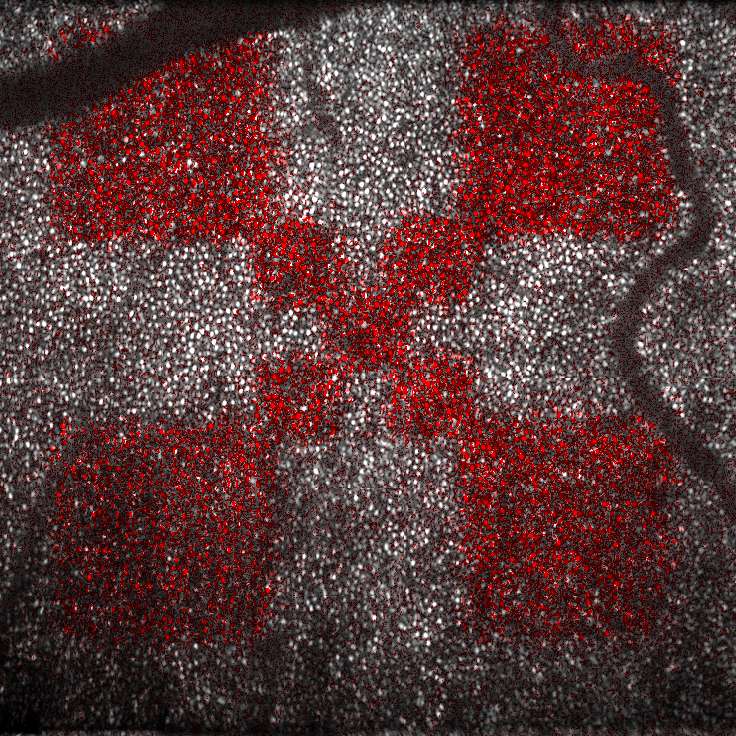
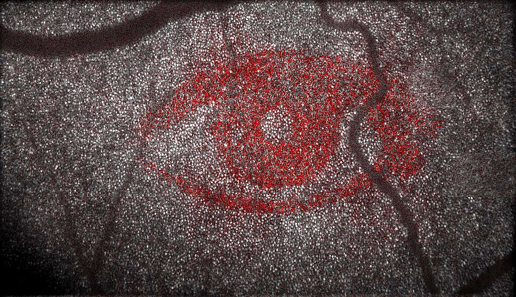
Messung der Reaktion von Photorezeptoren auf Licht
Aber auch noch wesentlich kleinere Veränderungen lassen sich mit Hilfe der phasensensitiven Holoskopie detektieren. Im Jahr 2015 ist es uns weltweit zum allerersten Mal gelungen, den Sehprozess in der Retina eines lebenden Menschen räumlich aufgelöst sichtbar zu machen. In der klassischen OCT verhindern Abbildungsfehler des Auges, dass einzelne Photorezeptoren dargestellt werden können. Durch eine numerische Aberrationskorrektur können in unseren Aufnahmen die Abbildungsfehler des Auges jedoch nachträglich kompensiert werden, so dass einzelne Photorezeptoren auflösbar werden. Kombiniert mit einer phasensensitiven Auswertung der komplexen OCT-Daten ermöglicht dies, die Reaktion einzelner Photorezeptoren auf einfallendes Licht direkt zu visualisieren

Das Team
Leitung
Gereon Hüttmann

Gebäude 81
,
Raum 69
gereon.huettmann(at)uni-luebeck.de
+49 451 3101 3206
Wissenschaftliche Mitarbeiter / Postdocs
Clara Pfäffle

AG Hüttmann
Gebäude 66
,
Raum 18.00
cl.pfaeffle(at)uni-luebeck.de
+49 451 3101 3296
Leo Puyo

AG Hüttmann
Gebäude 66
,
Raum 20.00
leo.puyo(at)uni-luebeck.de
+49 451 3101 3294
Doktoranden / Ingenieure
Jonas Franke-Duggen

AG Hüttmann
Gebäude 66
,
Raum 18.00
jon.franke(at)uni-luebeck.de
+49 451 3101 3296
Studierende und Hiwis
Baris Bargu

AG Hüttmann
Gebäude 66
,
Raum 17.00
baris.bargu(at)student.uni-luebeck.de
+49 451 3101 3234
Abdu Halowh

AG Hüttmann
Gebäude 66
,
Raum 17.00
abdu.halowh(at)student.uni-luebeck.de
+49 451 3101 3234


