Erforschung neuartiger Laserlichtquellen
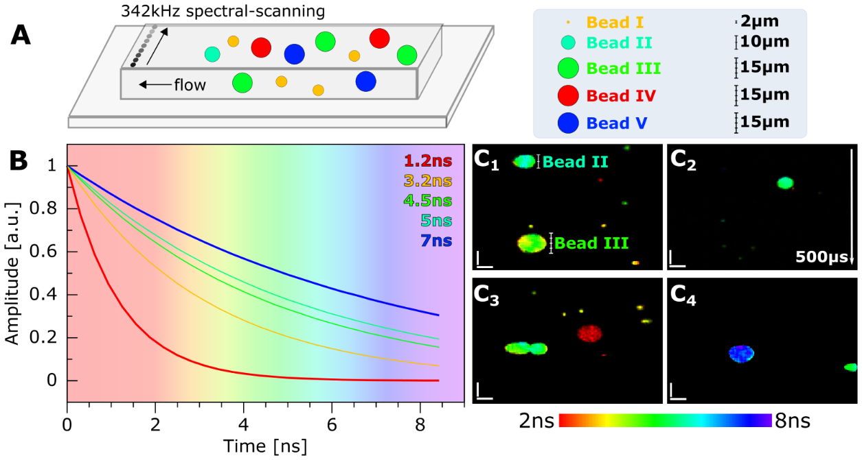
Die AG Karpf forscht an der Entwicklung neuartiger Laserlichtquellen und deren Einsatz in der biomedizinischen Bildgebung. Die jüngst entwickelte SLIDE-Mikroskopie ermöglicht schnellste Bildgebung in der nichtlinearen Zweiphotonenmikroskopie, bei der Bildwiederholraten von 2kHz und mehr erreicht werden. Solch schnelle Aufnahmegeschwindigkeiten gepaart mit der molekularen Spezifizität von Fluoreszenzmarkern und der hohen optischen Auflösung im sub-µm Bereich erlauben das Beobachten von schnellsten biologischen Vorgängen bis hinab in das Innere der Zellen. Eine Anwendung, die zukünftig mit dem SLIDE-Mikroskop an der Universität zu Lübeck verfolgt werden soll, ist die bildgebende Flusszytometrie, bei der binnen kürzester Zeit hunderte von Tausenden von Zellen bildgebend und multi-modal untersucht werden um extrem seltene Zellen, wie z.b. zirkulierende Tumorzellen, unter einer Vielzahl von gesunden Zellen zu diagnostizieren.

SLIDE Mikroskopie
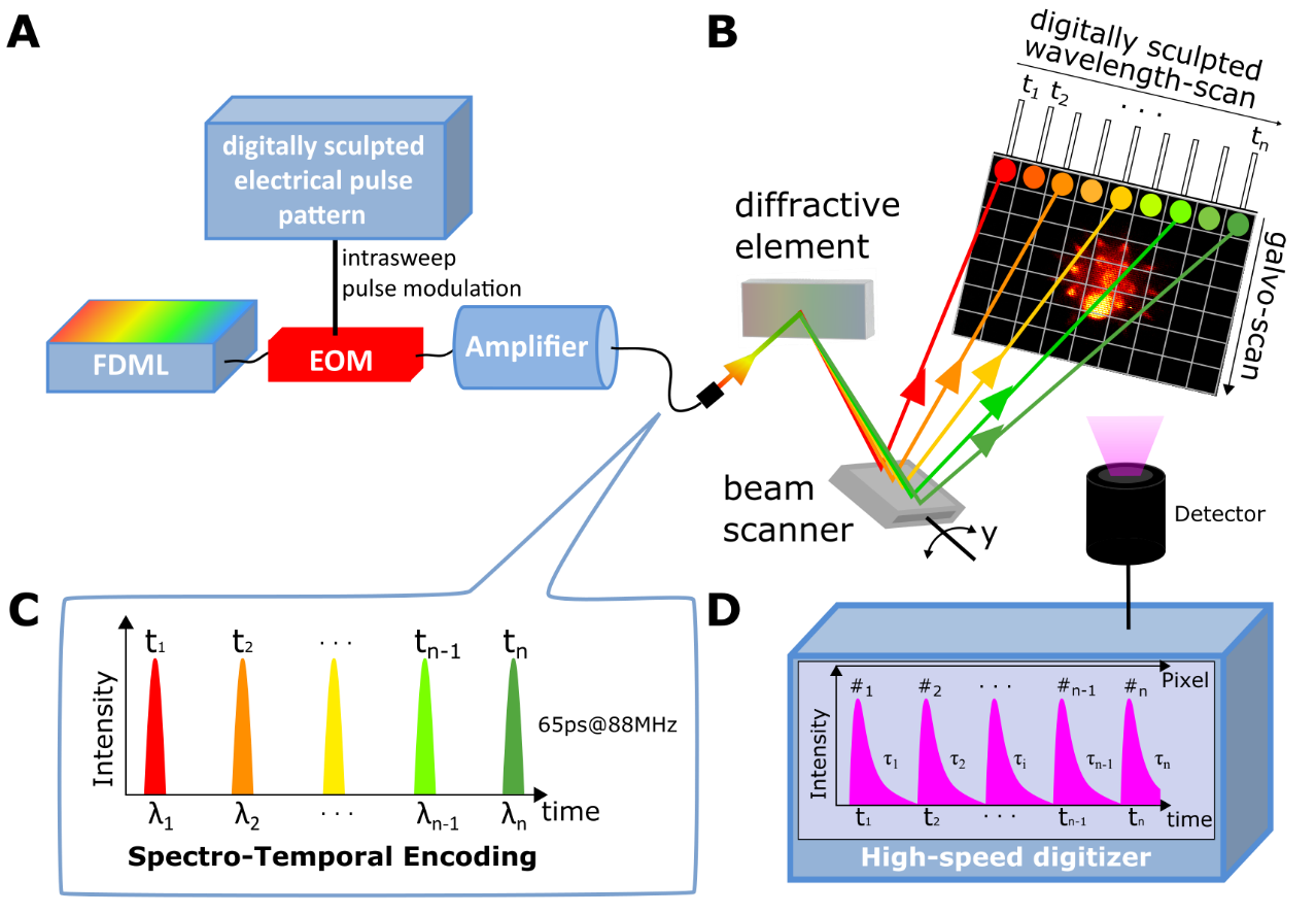
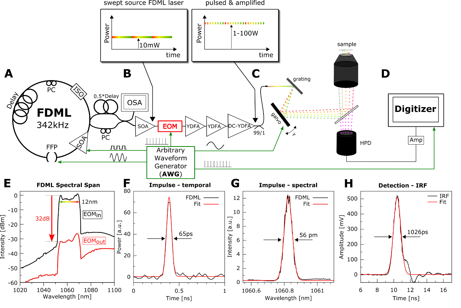
Das SLIDE Mikroskop wurde von Sebastian ‚Nino‘ Karpf an der UCLA, Kalifornien entwickelt, um schnellste biologische Vorgänge mittels nichtlinearer Zweiphotonenmikroskopie sichtbar zu machen. Moderne Zweiphotonenmikroskope verwenden galvanometrische Scanspiegel, die Zeilengeschwindigkeiten von ca. 20.000 Linien pro Sekunde erreichen. Diese Geschwindigkeiten sind limitiert durch das Trägheitsmoment der sich bewegenden Spiegel und können daher nicht beliebig erhöht werden. Will man zu schnelleren Scangeschwindigkeiten, muss man trägheitsfreie Scanverfahren verwenden. Das SLIDE-Mikroskop erreicht um Größenordnungen höhere Zeilenscangeschwindigkeiten indem es einen schnell wellenlängenabstimmbaren Laser einsetzt und diesen mittels einem Beugungsgitter ablenkt. Damit werden trägheitsfreie Scangeschwindigkeiten von MHz und mehr erreicht. Einen weiteren Vorteil bietet die schnelle Datenacquisition, die es ermöglicht auch transiente Fluoreszenzabklingkurven aufzunehmen und so mittels FLIM-Bildgebung auch Informationen aus der molekularen Umgebung der Fluoreszenzfarbstoffe zu extrahieren. Diese Multi-Modalität wird aus den selben Messdaten extrahiert, weshalb beide physikalischen Informationskanäle – TPEF und FLIM – mit der selben schnellen Bildwiederholrate von 2kHz und mehr aufgenommen werden.
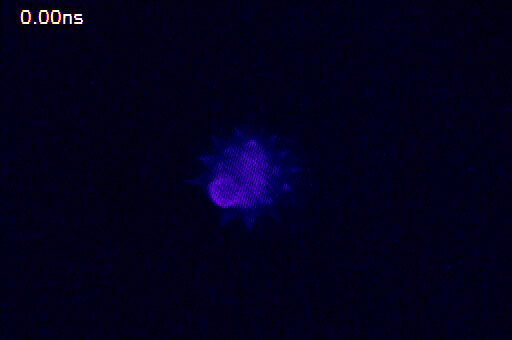
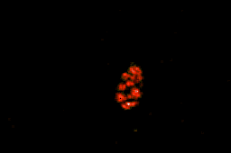
Diese Visualisierung veranschaulicht gut das Phänomen der Fluoreszenzlebenszeit, da zum Zeitpunkt 0ns der Anregungslichtimpuls auf die Probe geschickt wird und in der Folgenden Zeit unterschiedliche Teile der Polle das Leuchten (Fluoreszenz) anfangen und danach noch unterschiedlich lange Licht aussenden. Die Auswölbung unten links weist dabei die längste Fluoreszenzlebenszeit auf. Die Messung der Fluoreszenzlebenszeit erfolgte mittels SLIDE-Mikroskopie bei schneller Datenerfassung von 6,25 GSamples/s, einer Pixelrate von 176 MHz und einer Bildaufnahmegeschwindigkeit von 1ms pro Bild. Das hier dargestellte Video wurde aus 100 Aufnahmen gemittelt, was eine Gesamtaufnahmedauer von 100ms ergibt.
Falls Sie Interesse haben, bei diesem neuen Forschungsfeld mitzuwirken, dann schreiben Sie uns eine E-Mail. Wir suchen immer nach motivierten Bachelor- und Masterstudenten, die Neugier und Interesse an der biomedizinischen Forschung mitbringen und am Aufbau dieser neuen Technologie teilhaben möchten. Hier geht es zu den aktuellen Themen.

Gruppenleiter
Sebastian 'Nino' Karpf

Gebäude 81
,
Raum 71
sebastian.karpf(at)uni-luebeck.de
+49 0451 3101 3240
Doktoranden
Tonio Kutscher

AG Karpf
Gebäude Gartenhaus 58.600
,
Raum 11
t.kutscher(at)uni-luebeck.de
+49 451 3101 3291
Florian Sommer

AG Karpf
florian.sommer(at)student.uni-luebeck
+49 451 3101 3217
Christian Stock

AG Karpf
Gebäude Gartenhaus 58.600
,
Raum 11
ch.stock(at)uni-luebeck.de
+49 451 3101 3292
Stefan Reinhards

AG Karpf
Gebäude Gartenhaus 58.600
,
Raum 10
stefan.reinhards(at)uni-luebeck.de
+49 451 3101 3217
Matthea Thielking

AG Karpf
Gebäude Gartenhaus 58.600
,
Raum 10
m.thielking(at)uni-luebeck.de
+49 451 3101 3218
Gregory Berg

AG Karpf
Gebäude Gartenhaus 58.600
,
Raum 8
g.berg(at)uni-luebeck.de
+49 451 3101 3215
Studierende
Imke Klemme

AG Karpf
Gebäude 58.600
,
Raum 8
imke.klemme(at)stud.th-luebeck.de
+49 451 31013213


