Exploring the deep retina
The significance of oxidative stress in the pathogenesis of age-related macular degeneration
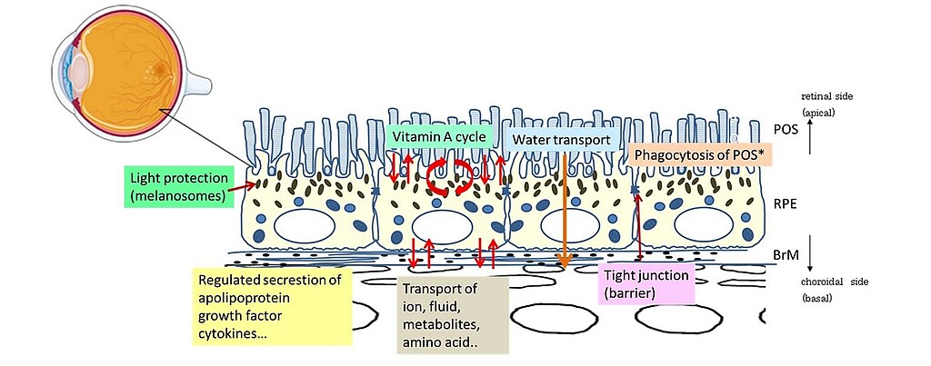
The retinal pigment epithelium (RPE) lies in the most outer layer of the retina between photoreceptor outer segments and the Bruch's membrane, and plays a key role in retinal physiology by forming the outer blood-retinal barrier and supporting the function of the neural retina (Fig.1). Its tight junctions function as a tight barrier strictly dividing the molecular circumstances in the choroid and the retina. The recent studies have suggested that oxidative stress in RPE cells leads to its functional disorders of RPE cells, such as disruption of the RPE junction integrity or lysosomal dysfunction. These RPE disorders lead to excess lipid accumulation inside and outside RPE cells, the induction of inflammatory cascades or formation of choroidal neovascularization, which are significantly relevant to the pathogenesis of age-related macular degeneration (AMD), one of the leading causes of blindness in western countries. Thus, protecting the RPE from oxidative stress is discussed as one of the therapeutic strategies of the treatment and prevention of AMD. If one can detect the cells or tissues suffering from oxidative stress-induced metabolic changes non-invasively, it must be absolutely a useful diagnostic tool for the prevention of many retinal diseases.

Group leader
Prof. Dr. Yoko Miura
Phone +49 (0)451 3101 3212
Fax +49 (0)451 3101 3204
yoko.miura@uni-luebeck.de


