UltraLas
Optical measurement techniques combined with ultrasound or laser tissue resection in neuro surgery for local detection of tissue boundaries, elasticity and vascular architecture
The UltraLas project is a joint project funded by the BMBF, which aims to improve tumor resection in neurosurgery. The Clinic for Neurosurgery of the University Medical Center Schleswig-Holstein, Campus Lübeck (headed by Dr. Matteo Mario Bonsanto, MD), the Institute for Biomedical Optics (BMO) of the University of Lübeck and the Medical Laser Center Lübeck (project management by Dr. Ralf Brinkmann) are involved in the project. Furthermore, the company Söring GmbH (Quickborn) and the company Asclepion Laser Technologies GmbH (Jena) are participating.
See also: https://mll-luebeck.com/oeffentliche-projekte/ultralas/
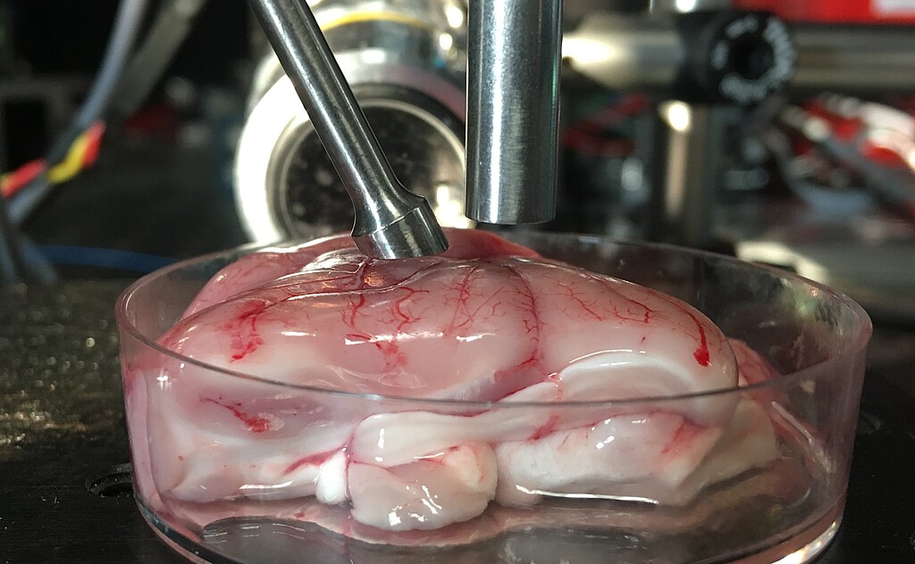
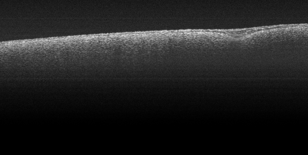
The aim of the project is to automatically detect the tumor size during the surgical procedure and to perform the resection of the diseased tissue as limited as possible to the tumor tissue. Since healthy and tumorous tissue differ optically, but especially haptically, the distinction between healthy and tumorous tissue has so far been made by manual palpation. This means that an experienced surgeon distinguishes the respective tissue structures based on their "firmness". The task of the BMO is to replace the subjective assessment by palpation with an objective, non-invasive measuring method. Specifically, elastography by means of optical coherence tomography (OCT) is to be used to measure the resection cavity and record tissue parameters.
In elastography, tissue is mechanically stressed, the tissue displacements in response to the stress are determined and the mechanical properties of the tissue are estimated from the displacements. If elastography is performed with optical coherence tomography, phase shifts resulting from the axial displacements of the tissue are usually measured by means of phase-sensitive OCT. There are several different approaches for mechanical loading or excitation of the tissue, including various dynamic and quasi-static methods. Within the project, different excitation sources and their suitability for the excitation of brain tissue will be investigated and the processes will be recorded using ultrafast OCT.