Optical microscopy has become an integral part of everyday medical practice. Whether in diagnostics, medical research or drug development, the high magnification achievable in microscopy allows insights into processes at the cellular level and thus provides intuitive and direct access to cause and effect in diseases. In addition to the high spatial resolution, time resolution is also an important aspect for the study of biological processes, which makes it possible to microscopically observe and physiologically interpret fast biodynamic processes such as cell movement in the blood flow or nerve action potential transmission.
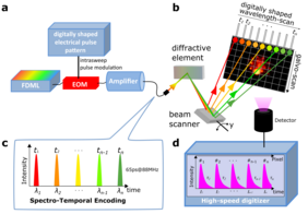
In a new publication in Nature Communications, Professor Sebastian 'Nino' Karpf of the Institute of Biomedical Optics (BMO) at the University of Lübeck reports on a new, ultra-fast microscope [1]. It is based on two-photon microscopy, which is a special form of fluorescence microscopy. It is similar to confocal microscopy, as it enables capturing high-resolution 3D images of tissue - an important aspect when shifting from research in the Petri dish to application in the body. The system developed by Prof. Karpf is based on a newly developed laser [2], a novel and extremely fast scanning method [3] as well as a highly efficient and fast light detection, which also enables fluorescence lifetime imaging [4]. The system is called SLIDE, an acronym for spectro-temporal laser imaging by diffractive excitation (SLIDE). It can acquire more than 2000 images per second, which is orders of magnitude faster than what was previously possible. Within local and international collaborations these high speeds are now applied in the detection of rare cells in flow cytometry, the imaging analysis of cells in liquid biopsy, the transmission of stimuli in the brain using neural imaging to study brain function and the study of immune cells in living organisms in situ.
The full text of the publication can be found here: https://www.nature.com/articles/s41467-020-15618-w
Original Publication: Karpf, S., Riche, C.T., Di Carlo, D. et al. Spectro-temporal encoded multiphoton microscopy and fluorescence lifetime imaging at kilohertz frame-rates. Nat Commun 11, 2062 (2020). https://doi.org/10.1038/s41467-020-15618-whttps>https://www.bmo.uni-luebeck.de/forschung/ag-karpf.htmlhttps
1. S. Karpf, C. T. Riche, D. Di Carlo, A. Goel, W. A. Zeiger, A. Suresh, C. Portera-Cailliau, and B. Jalali, "Spectro-temporal encoded multiphoton microscopy and fluorescence lifetime imaging at kilohertz frame-rates," Nature Communications, 11(1), (2020).
2. S. Karpf, and B. Jalali, "Fourier-domain mode-locked laser combined with a master-oscillator power amplifier architecture," Opt. Lett., 44(8), 1952 (2019).
3. Y. Jiang, S. Karpf, and B. Jalali, "Time-stretch LiDAR as a spectrally scanned time-of-flight ranging camera," Nature Photonics, 14(1), (2020).
4. M. Eibl, S. Karpf, D. Weng, H. Hakert, T. Pfeiffer, J. P. Kolb, and R. Huber, "Single pulse two photon fluorescence lifetime imaging (SP-FLIM) with MHz pixel rate," Biomed. Opt. Express, 8(7), 3132 (2017).


