Two-photon microscopy and fluorescence lifetime imaging of retinal cells
Two-photon microscopy (TPM) enables the measurement of tissue autofluorescence of thick tissues. Due to its special physical and optic properties, we may detect tissue autofluorescence only from the focused plain, so that the detailed investigation of intracellular autofluorescence with 3D and 4D imaging is enabled. Using this method, we investigate cellular autofluorescnece in different retinal cells, like RPE cells, photoreceptor outer segment, and retinal vascular endothelial cells under physiological and stress conditions (Fig. 1). Study of retinal cells with TPM provides us of the new insights on retinal cell pathology. Furthermore, we aim to realize in-vivo two-photon imaging using mouse model by cooperating with other research groups in our institute.
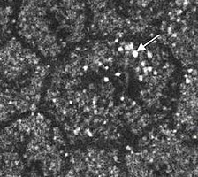
(Fig. 1) Two-photon microscopy of ex-vivo porcine RPE cells under oxidative stress. Very bright granular autofluorescence signals appear inside and around of RPE cell (arrow). (Miura et al. Two-photon microscopy and fluorescence lifetime imaging of retinal pigment epithelial cells under oxidative stress. Invest Ophthalmol Vis Sci. 2013 May 9;54(5):3366-77)
Fluorescence lifetime imaging microscopz (FLIM) is the technique that maps the spatial and temporal distribution of fluorescence lifetimes in microscopic images. Fluorescence lifetime is a fluorophore-intrinsic property. Since the status of fluorescence molecules changes depending on cell oxidative stress status and induced metabolic status, FLIM of the retina is discussed as a useful tool to detect retinal oxidative stress status and related metabolic changes. We study FLIM with TPM of the retinal tissues, in particular, of the RPE and photoreceptor outer segments under physiological and pathological conditions using ex-vivo tissues. We found a dramatic alteration of fluorescence lifetime of the RPE and POS under oxidative stress conditions (Fig. 2). We further investigate these fluorophores, with the purpose of the use of FLIM opthalmoscopy as an indicator for the development of retinal disease.
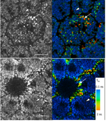
(Fig. 2) TPM (left) and FLIM (right) image of the ex-vivo RPE cells under oxidative stress. Very bright autofluorescence molecules appear inside (upper) and around (lower) of RPE cells (arrows). The fluorescence lifetime of these granular substances is much longer than the ones of melanosomes (arrow heads). (Miura et al. Two-photon microscopy and fluorescence lifetime imaging of retinal pigment epithelial cells under oxidative stress. Invest Ophthalmol Vis Sci. 2013 May 9;54(5):3366-77)

- Forschung
- AG Brinkmann
- AG Freidank
- AG Huber
- AG Hüttmann
- AG Karpf
- AG Linz
- AG Miura
- Member
- News and activities
- Research topics
- Minimal-invasice RPE thermal laser treatment
- Fluorescence lifetime imaging ophthalmoscopy (FLIO)
- Two-photon microscopy and fluorescence lifetime imaging of retinal cells
- Publications
- Conference presentations
- Open positions
- AG Rahmanzadeh
- AG Rahlves
- AG Vogel
- Emeritus Birngruber
- Publikationen
- Vortragsreihe Biomedizinische Optik
- Projekte und Drittmittel


