Optical measurement techniques combined with ultrasound and laser resection in neurosurgery for local detection of tissue boundaries, elasticity and vascular architecture (UltraLas)
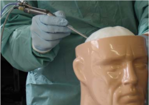
This BMBF-funded joint project is dedicated to improved tumour resection in neurosurgery. Optical measurement methods for recording tissue elasticity are to be combined with ultrasound and laser probes for tissue ablation.
In Germany, there are approximately 43,000 new oncological diseases of the central nervous system (CNS) every year, and this number will continue to rise in the future due to population development. Microsurgical resection is the standard treatment for the majority of tumours in the CNS. The survival rate depends on the extent of the resection. To date, the tumour margins to the intact tissue are still visible intraoperatively, but only with difficulty or with high technological effort. Further problems are the strongly fluctuating dissection rate when using ultrasound or laser instruments and the poor recognition of the underlying vascular courses. The consequences are a difficult choice of instrument parameters for the surgeon and intraoperative bleeding, which is currently mostly stopped with current flowing bipolar forceps. Disadvantages of this contact-based hemostasis include problems caused by tissue adhesion to the electrodes as well as an induced further difficulty in distinguishing the tumorous tissue by colour and feel. The overall goal of the collaborative research is therefore to improve the extent of resection during surgery by automatically detecting tumor characteristics and tumor edges. At the same time, the risk for the patient associated with the operation should be minimized, thus leading to an extended life span with a high quality of life.
In this project different methods for intraoperative in-vivo detection of tumor extent, vascular architecture and tumor elasticity in neurosurgery will be developed and evaluated. Several innovative photonic methods will be used for a fast and non-invasive measurement of the lesions on the one hand and in combination with therapeutic instruments on the other hand lead to an effective dissection and coagulation of the tumor tissue. Sub-goals are novel system solutions consisting of optical measuring methods in combination with laser and ultrasound instruments. Such a combination of different technologies combines intraoperative, comparatively inexpensive optomechanical tissue analysis with high local resolution and visualization of the deep-seated vascular architecture with an improved laser or ultrasound instrument. This novel optimization of the resection procedure enables a significantly improved tumor resection in clinical practice by supporting resection margin detection and optimized dissection performance. After completion of the project, the results will be transferred into marketable products.


