In this joint project funded by the Federal Ministry of Education and Research (BMBF), a three-dimensional, high-resolution real-time microscopy in neurosurgical operations is to be realized. The aim is to be able to remove tumours of the central nervous system more radically in the future and at the same time spare healthy brain tissue more effectively. Early recurrences should thus be prevented and patients should enjoy a better quality of life.
In addition to the BMO, the Department of Neurosurgery of the University Medical Center Schleswig-Holstein, Campus Lübeck, and the Medical Laser Center Lübeck (MLL) are involved. Funding by the German government in the Healthcare Industry Action Field of the German Health Research Framework Program amounts to 2.291 million EUR from October 2017 to September 2020.
In Germany, approximately 43,000 new oncological diseases of the central nervous system (CNS) are diagnosed every year. Microsurgical tumour resection of the CNS is often indicated. The more radically the neurosurgeon resects the tumour, the less frequently local tumour recurrences occur as a result. A problem here is the often poor recognition of tumor, vascular courses and tumor borders in contrast to healthy brain tissue. To visualize tumor and tumor borders, surgical microscopes and fluorescent dyes that accumulate in the tumor and make it visible under the surgical microscope with special filters are usually used, as well as neuronavigation. Further aids for tumor imaging are, if available, intraoperative ultrasound and intraoperative magnetic resonance imaging.
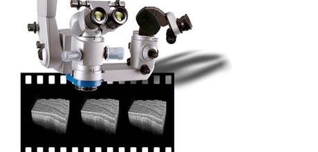
The aim of the Lübeck project is to compare the current intraoperative fluorescence procedure with two new, different intraoperative imaging procedures, namely with optical coherence tomography (OCT) (spectral domain OCT at 800 nanometers wavelength), which is already available in neurosurgery "as a prototype", and with an innovative high-frequency OCT (MHz-swept source OCT at 1300 nanometers wavelength), developed by Prof. Robert Huber, PI of the subproject of the Institute for Biomedical Optics.
The extremely fast, third-generation high-frequency OCT has the advantage that the entire resection area can be displayed in 3-D in real time. The higher wavelength also leads to a significantly lower tissue scattering, resulting in a higher tissue penetration depth. Furthermore, the optical imaging of blood vessels (angiography) is to be realized. In the clinical environment, both methods will be evaluated in terms of their sensitivity and specificity for tumor detection against fluorescence and validated in terms of tumor cell detection with the gold standard, histology.
This task is being pursued together with the Department of Neurosurgery and the Medical Laser Center Lübeck, which is also responsible for adapting the new OCT system to the surgical microscope. In addition, the consortium will also work out how 3D volume data can be optimally visualized in real time for the surgeon in order to continuously adapt the current tumor borders to the respective surgical step.
The aim of the project is to use this new technology to resect CNS tumours more radically in the future while sparing healthy brain tissue. The advantages: Prevention of early tumor relapses and improvement of the quality of life for patients. This consists in a reduced number of recurrent operations, which leads to fewer hospital stays; the prevention of neurological deficits makes it possible for patients to return to work or their social environment earlier.


